Blog

Big Endothelin-1 Biomarker Spotlight

Big Endothelin-1 (Big ET-1) is a protein that is mainly produced by vascular endothelial and smooth muscle cells and cardiomyocytes. Big ET-1 is a precursor to Endothelin (ET), the most potent vasoconstrictor known today. The half life of ET is less than one minute, whereas Big ET-1 is cleared more slowly. Big ET-1 can therefore be determined more easily, and has since been identified as a risk factor for a poor prognosis in patients with atrial fibrillation or coronary artery disease. A current study has shown that plasma concentrations of Big ET-1 is a valuable tool for risk stratification in patients with left ventricular non-compaction cardiomyopathy (LVNC). Other studies have also recognized the effectiveness of Big ET-1 as a predictor for numerous cardiovascular diseases.

Prognostic value of plasma big endothelin-1 in left ventricular non-compaction cardiomyopathy. Fan P, et al. Heart 2020;0:1–6. doi:10.1136/heartjnl-2020-317059

Plasma big endothelin-1 is an effective predictor for ventricular arrythmias and end-stage events in primary prevention implantable cardioverter- defibrillator indication patients. Li XY et al., 2020. J Geriatr Cardiol 28;17(7):427-433.

Plasma big endothelin-1 predicts new-onset atrial fibrillation after surgical septal myectomy in patients with hypertrophic cardiomyopathy. Song C et al., 2019. BMC Cardiovasc Disord 22;19(1):122.

Plasma level of big endothelin-1 predicts the prognosis in patients with hypertrophic cardiomyopathy. Wang Y et al., 2017. Int J Cardiol 15;243:283-289.


Related Products:

Big Endothelin-1 ELISA Assay Kit.
NT-proANP ELISA Assay Kit
NT-proCNP ELISA Assay Kit
NT-proBNP ELISA
BNP Fragment ELISA Assay Kit

If you have any questions regarding our Big Endothelin-1 ELISA Assay Kit or any other kits in the Cardiovascular Assay line, contact us here.

ELISpot Kits

Eagle Biosciences proudly offers a broad range of products in our extensive catalog. In addition to the well known ELISA format, we offer several assay kits in a enzyme-linked immunospot (ELISpot) assay format.

ELISA vs. ELISpot

ELISAs use antibodies to detect the presence and concentration of a protein in a liquid sample. In the most common form, antigens are applied to a stable surface (usually a microtiter plate). Then the corresponding antibody, linked with an enzyme, is added to form a complex. Finally, the enzyme’s substrate is added, which produces a measurable reaction, usually in the form of a color change.

ELISpots also utilize antibody-enzyme reactions. However, instead of measuring the concentration of protein in a sample, they measure the number of cells secreting the cytokine of interest in a sample. Antigens are applied to the plate surface, and when samples are added, cells settle on the bottom of the wells. The cytokines secreted by the cells are captured by the surrounding antigens. After the appropriate incubation time, the cells are removed and the secreted cytokine is detected using a detection antibody linked with an enzyme. The enzyme’s substrate is added, producing a reaction. The end result is visible spots on the on the surface of the plate. Each spot corresponds to an individual cell that was secreting the target protein.

The information provided by ELISpot kits is applicable to various fields of study including: transplantation, vaccine development, T-cell regulation analysis, autoimmune disease, cancer, allergy, and viral infections. These kits are highly sensitive and easy to use.

ELISpot Kit Procedure

Our available kits:

IFN gamma ELISpot Assay Kit

Mouse IFN gamma ELISpot Assay Kit

Rat IFN gamma ELISpot Assay Kit

TNF alpha ELISpot Assay Kit

Rat TNF alpha ELISpot Assay Kit

Contact us if you are looking for a specific ELISpot or if you have questions about any of our products.

Calprotectin ELISA

Does Human Milk Fortifier Affect Intestinal Inflammation in Preterm Infants?

Eagle Biosciences’ Calprotectin ELISA Assay Kit was used in a recent study dealing with inflammation of preterm infants in response to human milk fortifier. The Calprotectin ELISA Assay is part of our line of Gastrointestinal Assays which is a line of highly sensitive and specific kits used to detect the concentration of a variety of samples in serum, plasma, tissue, urine, and saliva.

Abstract


Background: Fecal calprotectin, a recognized marker of intestinal inflammation, is derived from neutrophil migration to a site of inflammation. Introduction of bovine-based human milk fortifier containing intact protein in preterm infants is associated with an increase in fecal calprotectin suggestive of intestinal inflammation. Newer fortifiers contain protein hydrolysates in place of intact protein.

Objective: To measure fecal calprotectin in human milk-fed preterm infants before and after human milk fortification using a fortifier containing hydrolyzed protein.

Methods: Serial stool samples were collected from 24 infants beginning at the first week to 60 days postnatal age. To compare the effect of human milk fortification, samples collected before and after fortification were compared. Infant demographics, diet, postnatal morbidities, and maternal characteristics were recorded.

Results: A total of 401 stool samples were collected from 24 study infants who had a birth weight of 993 ± 277 g (mean ± standard deviation), gestational age 27.5 ± 2.8 weeks, and fortifier initiation at 14 days. Median fecal calprotectin before and after fortification were similar. Calprotectin levels were not correlated with birth weight or gestational age but were inversely correlated with postnatal age (p = 0.005), use of fortifier (p < 0.001), receipt of antibiotics antenatally (p = 0.007) and postnatally (p = 0.008). After adjusting for postnatal age, calprotectin levels were significantly lower following receipt of fortifier (p < 0.001) and postnatal antibiotics (p < 0.001).

Conclusions: The feeding of protein hydrolysate-containing human milk fortifiers does not appear to be associated with increases in a marker of intestinal inflammation.

Click here for the full text.

Related Products

NGAL (Stool) ELISA Assay Kit
S100A8/A9 (MRP8/14, Calprotectin, Mouse/Rat) ELISA Kit
Stool Sample Collection Kit

COVID-19 IgG and IgA ELISA Feature in Recent Study Analyzing Antibody Response to SARS-CoV-2 Exposure and Symptom Onset

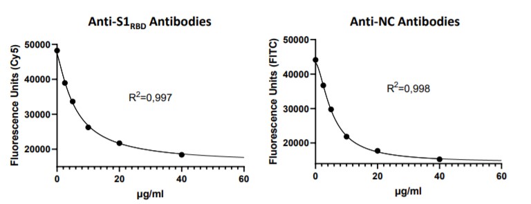
A study has been published in Viruses titled Persisting Neutralizing Activity to SARS-CoV-2 over Months in Sera of COVID-19 Patients by B, Flehming et al. This study takes a look at the long term presences of SARS-CoV-2 RNA and IgG and IgA antibodies in three patients diagnosed with COVID-19 over a six month period. This research utilized the COVID-19 Nucleocapsid IgG Quantitative ELISA Assay Kit and the Anti-SARS-CoV-2 IgA (S1 RBD) ELISA Assay offered by Eagle Biosciences. The results of this study indicate that immunity to the virus may persist for a couple of months after onset of symptoms. These findings are consistent with some other studies done with larger cohorts of COVID-19 patients, however similar longitudinal studies should be done in the future to corroborate these findings.

Abstract


The relationship between the nasopharyngeal virus load, IgA and IgG antibodies to both the S1-RBD-protein and the N-protein, as well as the neutralizing activity (NAbs) against SARS-CoV-2 in the blood of moderately afflicted COVID-19 patients, needs further longitudinal investigation. Several new serological methods to examine these parameters were developed, validated and applied in three patients of a family which underwent an ambulatory course of COVID-19 for six months. The virus load had almost completely disappeared after about four weeks. Serum IgA levels to the S1-RBD-protein and, to a lesser extent, to the N-protein, peaked about three weeks after clinical disease onset but declined soon thereafter. IgG levels rose continuously, reaching a plateau at approximately six weeks, and stayed elevated over the observation period. Virus-neutralizing activity reached a peak about 4 weeks after disease onset but dropped slowly. The longitudinal associations of virus neutralization and the serological immune response suggest immunity in patients even after a mild clinical course of COVID-19.

Click here for the full text.

To view Eagle Biosciences’ extensive collection of SARS-CoV-2 Assays and other products click here. 

Introducing the FGF-19 ELISA Assay Kit and the Mouse FGF-21 ELISA Kit. These kits are an extension of an expansive FGF Assay line of kits Eagle Biosciences already offers. Fibroblast Growth Factors are a family of cell signaling proteins that are involved in a wide variety of biological processes. FGF-19 and FGF-21 belong to a subfamily of these proteins that function as an important role in nutrient metabolism.

More about FGF-19:

The primary source of endocrine FGF-19 is the ileum, bile acids release into the intestine after a meal to induce expression of FGF-19. Circulating FGF-19 plays an important role in maintaining proper bile acid homeostasis. Several pharmacologic studies in hyperglycemic, obese animal models have shown that FGF-19 can improve metabolic rate and lower serum glucose and hepatic triglyceride and cholesterol levels. Like insulin, FGF-19 functions as postprandial hormone to govern hepatic protein synthesis, glycogen synthesis and gluconeogenesis, but does not stimulate lipogenesis.

More about FGF-21:

FGF-21 has been implicated in the regulation of lipid and glucose metabolism under fasting and ketotic conditions. In murine models, FGF-21 is predominantly expressed in liver, but it also expressed in adipose tissue and pancreatic β-cells. FGF-21 stimulates glucose uptake in adipocytes. It also protects animals from diet-induced obesity when overexpressed in transgenic mice and lowers blood glucose and triglyceride levels when administered to diabetic rodents. When administered daily for 6 weeks to diabetic rhesus monkeys, FGF-21 caused a dramatic decline in fasting plasma glucose, fructosamine, triglycerides, insulin, and glucagon. Furthermore, elevated plasma FGF-21 concentrations in humans appear to be related to the presence of hepatic and peripheral insulin resistance.

Related Products:

FGF-23 C-Terminal ELISA Kit

MedFrontier Intact FGF-23 Assay

FGF Basic ELISA Assay

Intact FGF-21 ELISA Kit

Total FGF-21 ELISA Assay

cTerminal FGF-21 ELISA Kit

N-Terminal FGF-21 ELISA Kit

iLite FGF-21 Assay Ready Cells

If you are looking for any other specific related products or if you have questions about our offerings, contacts us here.

Studies show that Angiopoietin-2 is a strong predictor in COVID-19 patients

Two research teams find that increased levels of the proinflammatory cytokine Angiopoietin-2 predicts transfer to the ICU and is responsible for hypercoagulation observed in critically ill COVID-patients.

Studies:

Angiopoietin-2 as a marker of endothelial activation is a good predictor factor for intensive care unit admission of COVID-19 patients. Smadja DM et al., Angiogenesis, 2020;1-10. Full text

Elevated Angiopoietin-2 inhibits thrombomodulin-mediated anticoagulation in critically ill COVID-19 patients. Hultstrom M et al., MedRxiv preprint server, 2021. Full text

Click here for a summary of the findings.

Background

SARS-CoV-2 is the causative agent of the coronavirus respiratory disease COVID-19. It has a diverse range of symptoms and may cause severe illness, in particular in patients with cardiovascular risk factors (1).

Endothelial damage and inflammation in SARS-CoV-2 infection

The inflammatory cytokine storm occurring in COVID-19 patients, leads to the recruitment of leukocytes which damage the capillary endothelium. The endothelial damage and inflammation in several organs in SARS-CoV-2 infection is a direct consequence of viral involvement and of the host inflammatory response (2).
Despite the routine thrombosis prophylaxis as standard of care treatment, the major COVID-19 complication is the hyperactivation of the coagulation system indicating a poor prognosis among COVID-19 patients in intensive care (3).

Angiopoietin-2 (ANG2) is a soluble marker of endothelial activation

Angiopoietin-2 is an angiogenesis regulator that can be rapidly released by the activated endothelium upon thrombin or inflammatory cytokines. ANG2 induces inflammation and vascular hyperpermeability and correlates with adverse outcomes in several critical care syndromes (4, 5).

Angiopoietin-2 is a crucial factor to predict transfer to the ICU

In COVID-19 patients, ANG2 was recently reported by Smadja and colleagues to be a relevant factor to predict transfer to the ICU as it was associated with poor lung compliance (6). Thus, showing that endothelial activation reinforces the hypothesis of a COVID-19-associated microvascular dysfunction.

Angiopoietin-2 inhibits anticoagulation in critically ill COVID-19 patients

Hulstrom and colleagues recently demonstrated that ANG2 levels in critically ill COVID-19 patients correlate with disease severity, hypercoagulation, and mortality. In addition, the researchers provided novel in vivo evidence for a direct role for ANG2 in coagulation through binding to and inhibition of thrombomodulin-mediated anticoagulation. The scientists suggest that inhibition of ANG2 might be beneficial for treating critically ill COVID-19 patients, as well as other patients with hypercoagulation (7).

About the Angiopoietin ELISA

  • Low sample volume – only 10µl needed
  • Assay range optimized for clinical samples
  • Ready to use standards and 2 controls included
  • Highly specific epitope mapped capture and detection antibodies

The human Angiopoietin-2 ELISA kit was developed and manufactured by Biomedica

Literature

  1. Clinical course and risk factors for mortality of adult inpatients with COVID-19 in Wuhan, China: a retrospective cohort study. Zhou F et al., Lancet, 2020; 395(10229):1054-1062.
  2. Endotheliitis in COVID-19. Varga S. Der Pathologe, 2020; 41(Suppl 2):99-102.
  3. COVID-19 and coagulation: bleeding and thrombotic manifestations of SARS-CoV-2 infection. Al-Samkari H et al., Blood, 2020:136, 489-500.
    Role of Angiopoietin-2 in Vascular Physiology and Pathophysiology. Akwii RG et al., Cells, 2019; 8(5): 471.
  4. Circulating angiopoietin-2 and the risk of mortality in patients with acute respiratory distress syndrome: a systematic review and meta-analysis of 10 prospective cohort studies. Li F et al., Therapeutic advances in respiratory disease, 2020; 14, 1753466620905274.
  5. Angiopoietin-2 as a marker of endothelial activation is a good predictor factor for intensive care unit admission of COVID-19 patients. Smadja DM et al., Angiogenesis, 2020;1-10.
  6. Elevated Angiopoietin-2 inhibits thrombomodulin-mediated anticoagulation in critically ill COVID-19 patients. Hultstrom M et al., MedRxiv preprint server, 2021.

Sclerostin expression in trabecular bone is down-regulated by osteoclasts

Scientific Reports

Bone tissues have trabecular bone with a high bone turnover rate and cortical bone with a low turnover. Expression of sclerostin by osteocytes works to inhibit bone formation. A study performed recently aimed to evaluate the relationship between the secretion of sclerostin by osteocytes and the secretion of leukemia inhibitory factor (LIF) by osteoclasts. It was found that LIF secretion effectively suppresses sclerostin expression and promotes bone formation.

Read the full article here.


Eagle Biosciences offers a sensitive and reliable assay for the detection of sclerostin in serum and plasma samples:

Sclerostin ELISA Assay Kit

Mouse LIF ELISA Assay Kit

Human LIFR ELISA Assay Kit

Free Soluble RANKL ELISA Assay Kit

If you are looking for any other specific Bone Monitoring related products or if you have questions about our offerings, contact us here.

EagleBio Endocrinology

Confirmation and Identification of Biomarkers Implicating Environmental Triggers in the Pathogenesis of Type 1 Diabetes

Frontiers in Immunology

Numerous ELISA Assay Kits from our endocrinology line were used in a recent study published out of the University of Nebraska Medical Center in Omaha, Nebraska. These kits were used to analyze blood plasma for biomarkers that could implicate environmental triggers for the pathogenesis of Type 1 Diabetes. These kits included;

Anti-GAD ELISA Assay Kit

Anti-IA2 ELISA Assay Kit

Anti-tTG IgA ELISA Assay Kit

High Sensitive Anti-TPO ELISA Assay Kit

Abstract


Multiple environmental triggers have been proposed to explain the increased incidence of type 1 diabetes (T1D). These include viral infections, microbiome disturbances, metabolic disorders, and vitamin D deficiency. Here, we used ELISA to examine blood plasma from juvenile T1D subjects and age-matched controls for the abundance of several circulating factors relevant to these hypotheses. We screened plasma for sCD14, mannose binding lectin (MBL), lipopolysaccharide binding protein (LBP), c-reactive protein (CRP), fatty acid binding protein 2 (FABP2), human growth hormone, leptin, total adiponectin, high molecular weight (HMW) adiponectin, total IgG, total IgA, total IgM, endotoxin core antibodies (EndoCAbs), 25(OH) vitamin D, vitamin D binding protein, IL-7, IL-10, IFN-γ, TNF-α, IL-17A, IL-18, and IL-18BPa. Subjects also were tested for prevalence of antibodies targeting adenovirus, parainfluenza 1/2/3, Coxsackievirus, cytomegalovirus, Epstein-Barr virus viral capsid antigen (EBV VCA), herpes simplex virus 1, and Saccharomyces cerevisiae. Finally, all subjects were screened for presence and abundance of autoantibodies targeting islet cell cytoplasmic proteins (ICA), glutamate decarboxylase 2 (GAD65), zinc transporter 8 (ZNT8), insulinoma antigen 2 (IA-2), tissue transglutaminase, and thyroid peroxidase, while β cell function was gauged by measuring c-peptide levels. We observed few differences between control and T1D subjects. Of these, we found elevated sCD14, IL-18BPa, and FABP2, and reduced total IgM. Female T1D subjects were notably elevated in CRP levels compared to control, while males were similar. T1D subjects also had significantly lower prevalence of EBV VCA antibodies compared to control. Lastly, we observed that c-peptide levels were significantly correlated with leptin levels among controls, but this relationship was not significant among T1D subjects. Alternatively, adiponectin levels were significantly correlated with c-peptide levels among T1D subjects, while controls showed no relationship between these two factors. Among T1D subjects, the highest c-peptide levels were associated with the lowest adiponectin levels, an indication of insulin resistance. In total, from our examination we found limited data that strongly support any of the hypotheses investigated. Rather, we observed an indication of unexplained monocyte/macrophage activation in T1D subjects judging from elevated levels of sCD14 and IL-18BPa. These observations were partnered with unique associations between adipokines and c-peptide levels among T1D subjects.

To learn more and read the full publication, click here.

For a full list of Eagle Bioscience’s Endocrinology line of ELISA kits click here.

Biomarker of urinary 5-HIAA as a valuable predictor of acute appendicitis

Science Direct

Acute appendicitis is one of the most common causes of severe acute abdominal pain globally. Increased levels of 5-Hydroxyindole acetic acid (5-HIAA), a metabolite of serotonin, in urine has been associated with acute appendicitis due to the densely packed serotonin-containing cells in the appendix. A study performed recently aimed to evaluate and determine the significance of 5-HIAA as a diagnostic marker.

Read the full article here.


Eagle Biosciences offers a sensitive and reliable assay for the detection of 5-HIAA in urine samples:

5-HIAA ELISA Assay Kit

Serotonin ELISA Assay Kit

Serotonin Ultrasensitive ELISA Assay Kit

If you are looking for any other specific 5-HIAA related product or if you have questions about our offerings, contact us here.

Studies Show that Neuropilin-1 Could Play Role in Transmission of COVID-19

Eagle News

Two Independent Research Teams Find Possible Link Between Neuropilin-1 and SARS-CoV-2 Transmission

Neuropilin-1 (NRP1) is an essential transmembrane cell surface receptor acting primarily as a co-receptor for various ligands (i.e. VEGF, Semaphorins). Due to alternative splicing or shedding, the extracellular region can be released into circulation as soluble Neuropilin-1. NRP1 functions in many key biological processes including the neuronal, cardiovascular and the immune system. The virus SARS-CoV-2 is the causative agent of the coronavirus disease COVID-19 NRP1 is expressed in multiple cell types in the body but occurs primarily on cells in the lung, nose and brain (i.e. respiratory and olfactory epithelium as well as in the CNS). SARS CoV 2 enters the host cells by its spike proteins mainly through its binding to the angiotensin converting enzyme 2 (ACE2).

Click here for summary of findings.

Studies:

Neuropilin-1 facilitates SARS-CoV-2 cell entry and provides a possible pathway into the central nervous system. Cantuti-Castelvetri L et al., Science 13 Nov, 2020; Vol. 370, Issue 6518, pp. 856-860. Full publication.

Neuropilin-1 is a host factor for SARS-CoV-2 infection. Daly JL et al., Science, 13 Nov 2020; Vol. 370, Issue 6518, pp. 861-865. Full publication.

About the Neuropilin-1 ELISA:

  • Only assay that measures free and ligand bound soluble NRP1
  • Low sample volume – only 10µl needed
  • Highly specific using epitope mapped antibodies
  • Rigorously validated according to international guidelines

Contact us for more information about this or our other SARS-CoV-2 related products.