The Eagle Bioscience’s Dopamine ELISA Assay Kit was utilized in a recent publications! The aim of the study was to investigate the potential of iron oxide nanoparticles produced using ascorbic acid (AA-IONPs) against Parkinson’s Disease. Check out the abstract and full text below!


Abstract

One of the most prevalent neurological movement diseases affecting the geriatric population globally is Parkinson’s disease (PD). Recent studies have highlighted the potency of biomolecules in the generation of nanomaterials and also over their impact on neuroprotection. The objective of this research was to investigate the potential of iron oxide nanoparticles produced using ascorbic acid (AA-IONPs) against PD. Numerous analytical methods including UV–Vis analysis, Fourier-Transform Infrared Spectroscopy (FTIR), dynamic light scattering (DLS), and electron microscopy (SEM, TEM), were used to analyze the produced AA-IONPs. Nitric oxide, prostaglandin E2, and inflammatory cytokines analyses such as IL-6 and IL-1 were employed to assess the neuroprotective effect of synthesized AA-IONPs on inflammatory agent lipopolysaccharides driven murine microglial BV2 cells. And also Parkinson-induced C57BL/6 mice were given the nanoparticle treatment to confirm the in vivo effects of the produced nanoparticles. Our characterization findings had demonstrated that AA-IONPs have a significant role in acting as an ideal nano drug and may have the ability to reduce inflammation in in vitro murine microglial BV2. The outcomes of in vivo tests conclusively show that AA-IONPs had reduced neuroinflammation and enhanced motor coordination in Parkinson’s disease-induced rats.

Li, L., Luo, P., Wu, S. et al. Deciphering the neuroprotective effect of ascorbic acid mediated synthesis of iron oxide nanoparticles against Parkinson’s disease: an in vitro and in vivo approach. Macromol. Res. 31, 949–960 (2023). https://doi.org/10.1007/s13233-023-00186-x


If you have any questions about this product or any of our other offerings contact us here.


The Biomedica Human IL-6 High Sensitive ELISA Assay Kit was highlighted in a recent publication that explored factors associated with incident vertebral fractures in glucocorticoid-treated Duchenne muscular dystrophy. Check out the abstract and full text below!


Abstract

Purpose: Prevention of fractures is an unmet need in glucocorticoid (GC)-treated Duchenne muscular dystrophy. This study explored factors associated with incident vertebral fractures (VFs) to inform future fracture prevention efforts.

Methods: VFs were evaluated prospectively at study baseline and 12 months on lateral spine radiographs in participants aged 4 to 25 years with Duchenne muscular dystrophy. Clinical factors were analyzed for their association with the change in Spinal Deformity Index (sum of the Genant-defined VF grades from T4 to L4) between baseline and 12 months.

Results: Thirty-eight males were evaluated (mean ± SD age at baseline 11.0 ± 3.6 years; mean ± SD GC duration at baseline 4.1 ± 3.1 years; 74% ambulatory). Nine of 38 participants (24%) had 17 incident VFs, of which 3/17 VFs (18%) were moderate/severe. Participants with 12-month incident VF had lower mean ± SD baseline lumbar spine areal bone mineral density Z-scores (-2.9 ± 1.0 vs -1.9 ± 1.1; P = .049) and lower total body less head areal bone mineral density Z-scores (-3.1 ± 1.2 vs -1.6 ± 1.7; P = .036). Multivariable linear regression showed that at least 1 VF at baseline (P < .001), a higher number of antecedent non-VF (P < .001), and greater bone age delay at baseline (P = .027) were significant predictors of an increase in the Spinal Deformity Index from baseline to 12 months.

Conclusion: The observation that ≥ 1 prevalent VF and/or non-VF were the strongest predictors of incident VFs at 12 months supports the need for prevention of first fractures in this high-risk setting. Bone age delay, a marker of GC exposure, may assist in the prioritization of patients in efforts to prevent first fractures.

Keywords: Duchenne muscular dystrophy; bone fragility; glucocorticoids; incident fractures; osteoporosis; vertebral fractures.

Risk Factors Associated with Incident Vertebral Fractures in Steroid-treated Males with Duchenne Muscular Dystrophy . Phung K, McAdam L, Ma J, McMillan HJ, Jackowski S, Scharke M, Matzinger MA, Shenouda N, Koujok K, Jaremko JL, Wilson N, Walker S, Hartigan C, Khan N, Page M, Robinson ME, Saleh DS, Smit K, Rauch F, Siminoski K, Ward LMJ Clin Endocrinol Metab. 2023 Aug 23:dgad435. doi: 10.1210/clinem/dgad435. Epub ahead of print.PMID: 37610420


If you have any questions about this product or any of our other offerings, contact us here.

What is it AgarSqueezer intended for?

AgarSqueezer is a microscope slide chamber equipped with a molded agar-based compression system. Use it to assess cell response to short and long-term mechanical confinement within a physiological rigidity range.

It is very helpful if you want to analyze how your cells will react if you squeeze them for a prolonged period. Or if you want to study how mechanical confinement affects drug cell resistance. And if you want to perform immunostaining in situ.


Kit Description:

You can choose among 2 kits:

  • The “1 AgarSqueezer kit” contains 1 AgarSqueezer device
  • The “2 AgarSqueezers kit” contains 2 AgarSqueezers devices.

In addition to the device, each kit contains also:

  • 1 x insert to hold up to 2 AgarSqueezers on the microscope stage
  • 1 x 16G flat cut needle to make holes in the agar gel, facilitating diffusion of culture medium or drugs during the experiment
  • 1 x 20G flat cut needle (same)

For users who also need a wafer to mold agarose, contact us to learn about the four different heights we offer.


Features

  1. Tunable stiffness in a physiological range [1-150] kPa – Use of agarose as a cheap and biocompatible polymer.
  2. Open access to the reservoir – Possibility to add drugs, and reagents. Easy medium renewal.
  3. Autoclavable and reusable systems.
  4. Compatibility with multiple microscopy techniques – Confocal, spinning, super-resolution. Open access for microscope objectives. Use of optical glass coverslip to make cells grow.
  5. Easy to recover coverslip with cells for subsequent molecular analysis – FACS, qPCR, Western-Blot, Immunoflourescence (possible in situ).
  6. Long-term analysis of the cell adaptation to confinement – Up to several days, for time-lapse studies.
  7. Study of the specific impact of mechanical loads on the biology of cells – Gas permeability of the system allows to get rid of the hypoxia conditions.
  8. Easy to assemble and disassemble the system

For more information about this product or any others from the Microscopy line, contact us here.

Eagle Biosciences will be at ASN Kidney Week in Philadelphia, PA!

Eagle Biosciences will be at ASN Kidney Week next week, Thursday, November 2 to Sunday, November 5, at the Pennsylvania Convention Center. Stop by booth #1642 to learn more about Intact FGF23, Fetuin A, and other assays that could help you with your kidney related research! We’ll be there to answer any questions you may have, or stop and say hi! We love seeing our customers!


Product Highlights

Intact FGF23: This CLEIA ELISA is a unique and highly accurate assay for the measurement of FGF23, which has been linked to impaired renal function.

Fetuin A (PTM): This ELISA is a unique immunoassay for the measurement of Fetuin-A with specific post translational modifications in urine samples. This biomarker has shown to be an early indicator of diabetic kidney disease (DKD).


If you have questions about either of these products or any of our other offerings, contact us here.

Eagle Biosciences is excited to bring you a wide array of more complement assays!

The complement system is an essential part of the immune system in the human body, playing a crucial role in defending against infections, clearing cellular debris, and promoting inflammation. It consists of a complex network of proteins and molecules that work together to enhance the immune response. The complement system can be activated through three main pathways, the classical pathway, the alternative pathway, the lectin pathway. The complement system is tightly regulated to prevent excessive immune responses and potential damage to host tissues. Various regulatory proteins are involved in controlling the activation and amplification of the complement cascade to maintain a delicate balance between defense and protection.


New Complement Assays

Rat Classical Complement Pathway ELISA Assay Kit
Rat Lectin Complement Pathway ELISA Assay Kit
Rat Alternative Complement Pathway ELISA Assay Kit
Mouse Classical Complement Pathway ELISA Assay Kit
Mouse Alternative Complement Pathway ELISA Assay Kit
Pig Classical Complement Pathway ELISA Assay Kit
Pig Lectin Complement Pathway ELISA Assay Kit
Pig Alternative Complement Pathway ELISA Assay Kit
Pig Complement Pathway ELISA Assay Kits
MASP1/C1-INH Complex ELISA Assay Kit
C3d ELISA Assay Kit
MASP-2 ELISA Assay Kit
Complement Factor H ELISA Assay Kit
Complement Factor D ELISA Assay Kit
Complement Factor I ELISA Assay Kit
Collectin-10 ELISA Assay Kit
Factor B ELISA Assay Kit
sCD59 ELISA Assay Kit
C1s/C1-INH Complex ELISA Assay Kit
Rat Terminal Complement Complex (TCC) ELISA Assay Kit
Mouse C3 ELISA Assay Kit
Mouse C1q ELISA Assay Kit
Mouse C3b ELISA Assay Kit
Mouse C4 ELISA Assay Kit


Find the complete complement product catalog here. If you have any questions about any of these products or our other offerings, contact us here.

Eagle Biosciences will be at AACC in Anaheim, California!

This year AACC is in Anaheim at the Anaheim Convention Center, Sunday July 23th – Thursday July 27th. We will be at booth #1147 from July 25th to July 27th! Come by to learn about the GA-Map Dybiosis Test Lx and more assays that could help you with your microbiome or other research! We will be there to answer any questions you may have, or just stop by and say hi! We love seeing our customers!


Product Highlights

GA-Map Dysbiosis Test Lx: The first and only standardized solution for microbiome profiling! The GA-Map Dysbiosis Test Lx is a simple multiplex stool assay that maps the intestinal microbiota profile for a selected set of bacteria. The GA-map® platform uses probes that target variable regions (V3 to V7) of the bacterial 16S rRNA gene to characterize and identify bacteria present. The targets are identified in a molecular multiplex assay that utilizes the Single Nucleotide Primer Extension (SNuPE) technology patented by Professor Knut Rudi (US6617138). A unique algorithm takes advantage of all the data generated by the detection of the SnuPE products to determine dysbiosis level in the sample. The algorithm is incorporated in the GA-map® Dysbiosis Analyzer software that accompanies the test.

GA-map® Dysbiosis Test Lx Procedure Quick Guide

GA-map Dysbiosis Test


If you have any other questions about these products or our other offerings, contact us here.


Eagle Bioscience’s is excited to highlight the Alpha Synuclein Preformed Fibrils (Type 1) from StressMarq!


About the Alpha Synuclein Pre-Formed Fibrils (Type 1)

StressMarq’s Alpha Synuclein Pre-formed Fibrils (Type 1) are produced with low endotoxin levels, making them the suitable choice for both in-vivo and in-vitro work. Alpha Synuclein PFFs seed the formation of new fibrils from active alpha synuclein monomers, and can be used to induce endogenous alpha synuclein phosphorylation and subsequent Lewy body inclusion formation in neuronal cell culture or for in vitro oligomerization studies.


Background

Alpha-Synuclein (SNCA) is expressed predominantly in the brain, where it is concentrated in presynaptic nerve terminals1. Alpha-synuclein is highly expressed in the mitochondria of the olfactory bulb, hippocampus, striatum and thalamus2. Functionally, it has been shown to significantly interact with tubulin3, and may serve as a potential microtubule-associated protein. It has also been found to be essential for normal development of the cognitive functions; inactivation may lead to impaired spatial learning and working memory4. SNCA fibrillar aggregates represent the major non A-beta component of Alzheimers disease amyloid plaque, and a major component of Lewy body inclusions, and Parkinson’s disease. Parkinson’s disease (PD) is a common neurodegenerative disorder characterized by the progressive accumulation in selected neurons of protein inclusions containing alpha-synuclein and ubiquitin5, 6.

ATTO633 fluorescently-labelled alpha synuclein PFFs (SPR-322) were taken up, transported into the soma, and induced alpha synuclein aggregation in mouse neurocortical primary cells. (A) Neurites filled with fluorescently-labelled alpha synuclein seeds in a microfluidic co-culture system after 24 hours. (B) Alpha synuclein seeds within the soma and neurites of mouse neurocortical primary cells after 24 hours. Experiment and imaging courtesy of Cellectricon.

References

  1. “Genetics Home Reference: SNCA”. US National Library of Medicine. (2013).
  2. Zhang L., et al. (2008) Brain Res. 1244: 40-52.
  3. Alim M.A., et al. (2002) J Biol Chem. 277(3): 2112-2117.
  4. Kokhan V.S., Afanasyeva M.A., Van’kin G. (2012) Behav. Brain. Res. 231(1): 226-230.
  5. Spillantini M.G., et al. (1997) Nature. 388(6645): 839-840.
  6. Mezey E., et al. (1998) Nat Med. 4(7): 755-757.

The Alpha Synuclein Preformed Fibrils (Type 1) were utilized in the following studies:


If you have any questions about this product or any of our other offerings, contact us here.

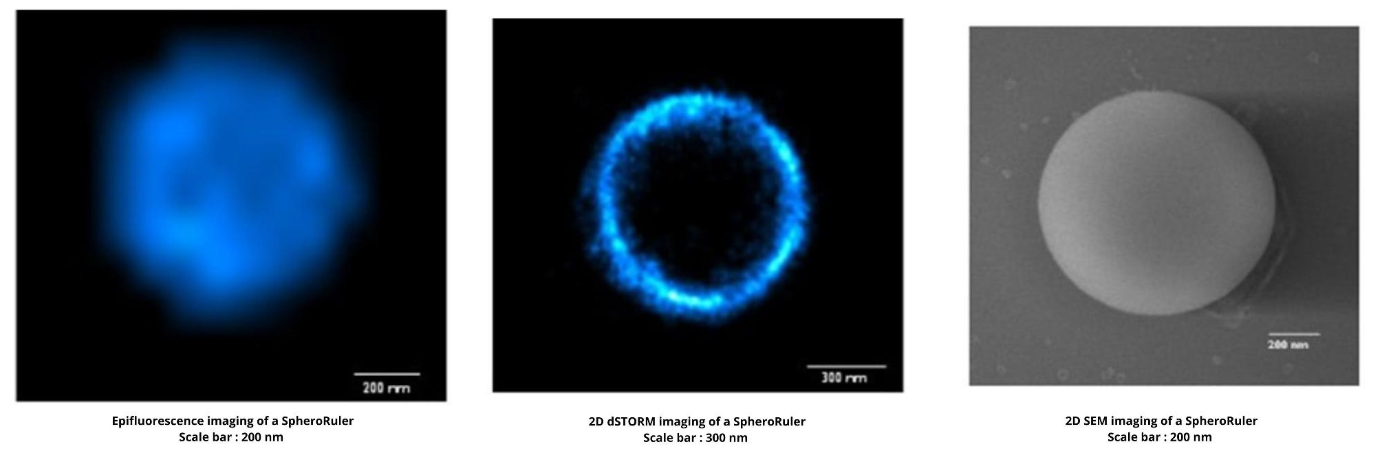
What is it SpheroRuler intended for?

SpheroRuler is a monodispersed suspension of 1µm diameter spheres coated with 647-fluorophores giving a stable blinking in SMLM microscopy. Their consistent size and geometry make them very practical and reliable standards to assess the accuracy of your x-y or z measurements, 3D reconstruction methods or your image quality. Monodispersed and immobile in solution, they can also be used as rulers, for drift correction or as guides to help localize features on your biological samples.

SpheroRuler is great for dSTORM calibration experiments because you can use it on your preferred support and buffer. And because you get confident with the new biological structure to image. SpheroRuler also produces simple shapes that are easy to study when you are a dSTORM beginner!


Features

  1. Stable Blinking – Suited for use in SMLM microscopy
  2. Consistent Size – Thoroughly characterized by electron microscopy
  3. Spherical geometry – with sharp and thin edges
  4. Immobile – Suitable for drift correction applications
  5. Bio-compatible – Resuspended in an aqueous buffer and usable along biological samples such as cells or tissue sections
  6. Easy-to-Spot – A high fluorescence intensity for practical use as demo or training tools


For more information about this product or any others from the Microscopy line, contact us here.

The Eagle Bioscience’s 25-OH Vitamin D ELISA Assay Kit has been used in a number of recent publications! These studies range from exploring the effects of vitamin D on cellular responses, molecular immunity, and mycobacterial killing in cattle, to the role of vitamin D, DKK1, hepcidin, and other oxidative stress biomarkers in type 2 diabetes mellitus patients. Check them all out below!


Flores Villalva, Susana. “The Effects of Vitamin D on the Cellular Responses, Molecular Immunity, and Mycobacterial Killing in Cattle.” University College Dublin. School of Agriculture and Food Science, 2022.

Kamel, Amira A., et al. “The Role of Vitamin D, DKK1, Hepcidin and Oxidative Stress Biomarkers in Type 2 Diabetes Mellitus Patients with and without Diabetic Nephropathy.” The Egyptian Journal of Hospital Medicine, vol. 89, no. 2, 2022, pp. 7137–7146.

Blakely, L.P., et al. “Effect of Vitamin D Source and Amount on Vitamin D Status and Response to Endotoxin Challenge.” Journal of Dairy Science, vol. 106, no. 2, 2023, pp. 912–926.

Fernández-Lázaro, Diego, et al. “25-Hydroxyvitamin D Serum Levels Linked to Single Nucleotide Polymorphisms (Snps) (RS2228570, RS2282679, rs10741657) in Skeletal Muscle Aging in Institutionalized Elderly Men Not Supplemented with Vitamin D.” International Journal of Molecular Sciences, vol. 23, no. 19, 2022, p. 11846.

Kumar, Abhai, et al. “Vitamin D and Inflammatory Cytokines Association in Mild Cognitive Impaired Subjects.” Neuroscience Letters, vol. 795, 2023, p. 137044.

Stenhouse, Claire, et al. “Uptake of Phosphate, Calcium, and Vitamin D by the Pregnant Uterus of Sheep in Late Gestation: Regulation by Chorionic Somatomammotropin Hormone.” International Journal of Molecular Sciences, vol. 23, no. 14, 2022, p. 7795.


If you have any questions about our 25-OH Vitamin D ELISA Kit or any of our other offerings, contact us here.

The MedFrontier Intact FGF-23 Assay is developed and manufactured by Minaris Medical! This assay kit is a reliable test that provides a simple and quick assay procedure that yields a broad dynamic range.


What is FGF-23?

FGF-23 (Fibroblast Growth Factor 23) is a protein belonging to the fibroblast growth factor family. FGF-23 is involved in the regulation of phosphorus metabolism. FGF-23 has a molecular weight of approximately 32 kDa. FGF-23 is produced in bone cells. In vivo, FGF-23 is secreted into circulation. A full-length active FGF-23 protein may undergo proteolytic cleavage to generate an inactive c-terminal fragment. The MedFrontier Intact FGF-23 Assay measures ONLY the full-length active form (intact form).

Why Measure FGF-23?

Phosphate plays a critical role in the formation and growth of bones in children and maintaining strength in adults. If there is an imbalance of FGF-23 within the bloodstream, it can cause hyper- and hypophosphatemia. These conditions have been linked to Rickets, osteomalacia, and impaired renal function to name a few.


About the MedFrontier Intact FGF-23 Assay Kit

This is a sandwich Chemiluminescence enzyme immunoassay (CLEIA) kit by using two anti-human FGF-23 mouse monoclonal antibodies. When serum is added to a plate well coated with anti-human FGF-23 mouse monoclonal antibodies, FGF-23 is captured by the immobilized antibodies (1st reaction). After the 1st reaction, the plate is washed. Then, ALP-labeled anti-human FGF-23 mouse monoclonal 2nd antibodies against FGF-23 react to FGF-23 antigens captured by the immobilized antibodies (2nd reaction). After the 2nd reaction, the plate is washed and light detection is performed after adding the luminescence reagent. Each active well of the plate is measured using a luminescence microplate reader and relative light units (RLU) are obtained. The concentration of FGF-23 in serum is calculated with a standard curve generated using the FGF-23 Standard 1 to 6.

Check out the product flyer to learn more!


If you have any questions about this kit or any of our other offerings, contact us here.