We, at Eagle Biosciences, Inc. are rapidly expanding our selection of Coronavirus Assays by welcoming two new research kits! With the ongoing pandemic, it’s more important now than ever that we continue to offer the highest quality assays to our customers. Here are our newest additions;
Anti-SARS-CoV-2 S1 (RBD) IgG ELISA Assay
Catalog Number: E111
The Anti-SARS-CoV-2 S1 (RBD) IgG ELISA Assay Kit is manufactured in Germany by Mediagnost. This assay is a highly specific enzyme immunoassay for the detection of IgG antibodies directed against SARS-CoV-2-S1 Receptor Binding Domain (RBD) in human blood. In this assay the recombinant Receptor Binding Domain (RBD) of SARS-CoV-2 S1 spike protein, which binds the ACE2 receptor, is used. The use of RBD increases the specificity of the assay since the domain is identical with SARS-CoV but not with MERS-CoV for example. Antibodies directed against the RBD neutralize both virus strains SARS-CoV and SARS-CoV-2.
Size: 1×96 wells
Incubation Time: 2 hours 40 minutes
Sample Type: Serum and Plasma
Sample Size: 100 µl
Controls Included
To learn more about the Anti-SARS-CoV-2 S1 (RBD) IgG ELISA Assay click here*
Anti-SARS-CoV-2 (S1, S2, N) IgG ELISA Assay Kit
Catalog Number: 3940
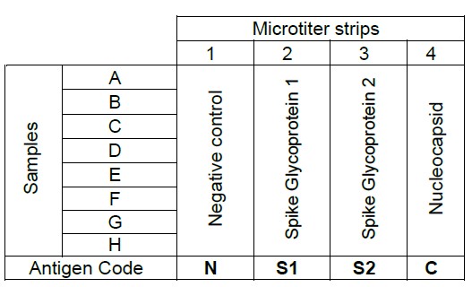
The Anti-SARS-CoV-2 (S1, S2, N) IgG ELISA Assay Kit is manufactured in Germany by Generic Assays. This assay is a module based Enzyme Immunoassay for the confirmation of positive IgG antibodies against SARS coronavirus 2 (SARS-CoV-2) in the first screening. The Anti-SARS-CoV-2 (S1, S2, N) IgG ELISA Assay Kit determines the specificity of antibodies against the main immunodominant antigens (Spike Glycoprotein 1, Spike Glycoprotein 2, Nucleocapsid) of SARS-CoV-2 in human serum or plasma. This test kit consists of modules separately coated with the major antigens of the virus as seen in the illistration below:
Size: 96 wells (24 samples x 4)
Incubation Time: 2 hours
Sample Type: Serum or Plasma
Number Of Tests Per Kit: 24 (22 samples + controls)
Sample Size: 50 µl
Controls Included
To learn more about the Anti-SARS-CoV-2 (S1, S2, N) IgG ELISA Assay Kit click here*
Check out our entire Coronavirus Series here or contact us with any questions or inquires
*These kits are for research use only and should not be used for diagnostic procedures.