Eagle Biosciences is thrilled to introduce the Anti-MyD88 L265P Antibody to our product lineup! This powerful tool can significantly enhance your lymphoma research.


Application

MyD88 L265P is a prevalent driver mutation in B-cell lymphomas, contributing to oncogenic signaling by activating BTK, HCK, NFkB, and JAK-STAT3 pathways. This mutation is linked to a poor prognosis and is frequently observed in:

  • 90% of Waldenström’s macroglobulinemia (WM) cases
  • 50-80% of IgM monoclonal gammopathy of undetermined significance (IgM-MGUS) cases
  • 29% of diffuse large B-cell lymphomas (DLBCL) cases

WM and the aggressive activated B-cell-like (ABC) subtype of DLBCL remain incurable, making the tumor-specific MyD88 L265P mutation a valuable prognostic marker and a promising target for novel therapies.

Background

The MyD88 gene encodes a cytosolic adapter protein vital for both innate and adaptive immune responses. This protein acts as a crucial signal transducer in the interleukin-1 and Toll-like receptor signaling pathways, which regulate the activation of numerous proinflammatory genes. The protein includes an N-terminal death domain and a C-terminal Toll-interleukin1 receptor domain. Patients with defects in this gene are more susceptible to pyogenic bacterial infections. Multiple transcript variants result from alternative splicing.


If you have any questions about this product or any of our other offerings, contact us here.

Eagle Biosciences is thrilled to announce the expansion of our product line with several innovative new Host Cell Protein Detection Kits! These cutting-edge assays enhance our comprehensive range of Host Cell Protein Detection Kits, offering more precise and efficient solutions for your research needs. These additions reflect our ongoing commitment to providing high-quality, reliable tools for the biotechnology and life sciences communities. Discover our complete line of Host Cell Protein Detection Kits here.


Explore Our New Kits:


For more information or any inquiries about our products, please contact us here.

How does it work?

GlowMito provides a non-toxic solution to quickly visualize the entire mitochondrial network in live samples.

GlowMito quickly penetrates cells and produces a bright & stable red labeling of mitochondria without inducing cell toxicity or altering mitochondrial functions. produces a bright, red labeling of the entire mitochondrial network, including those with reduced potential. It is perfectly suitable for:

  • Live imaging: study of mitochondrial dynamics (velocity, localization), structural changes, multiplexing (potentiometric dyes, calcium signaling probes, etc)
  • Downstream analysis: flow cytometry, oxygraphy, etc

We do not recommend its use to measure mitochondrial mass or volume density.


Simplified Protocol


Frequently Asked Questions

How was mitochondrial specificity of GlowMito verified?
The strong ability of GlowMito to specifically target mitochondria has been meticulously checked by co-localization studies. More generally, the ability of lipophilic cations to specifically target mitochondria is already well-established and conjugating molecules to lipophilic cations is a commonly used method to develop mitochondria-targeted compounds.

Which assays can GlowMito be used for?
GlowMito produces a bright, red labeling of the entire mitochondrial network, including those with reduced potential. It is perfectly suitable for:

  • Live imaging: study of mitochondrial dynamics (velocity, localization), structural changes, multiplexing (potentiometric dyes, calcium signaling probes, etc)
  • Downstream analysis: flow cytometry, oxygraphy, etc

We do not recommend its use to measure mitochondrial mass or volume density.

Which types of samples has GlowMito been used with?
So far, GlowMito has been successfully used to label mitochondria in the following biological samples:

  • Human cells: HEK293, HeLa, MCF-7, MDA-MB-231, HMLE, UACC-62, U2-OS, Gli36, HAEC, SH-SY5Y, A-172, A549, patient-derived skeletal muscle cells & primary smooth muscle cells
  • Monkey cells: COS-7
  • Mice cells: primary cortical neurons
  • Tissues: hiPSC-derived heart tissues & mice isolated pressurized blood vessels
  • Parasitic protists: Trichomonas Vaginalis

GlowMito showed no internalization in yeast cells


For more information about this product or any others from the Microscopy line, contact us here.

What is it intended for?

DNAbsolute is composed of a ionic liquid that has a selective and high affinity for DNA strands. When mixed with your sample, the DNAbsolute reagent will directly bind DNA and precipitate it without the need for RNase treatment, protein precipitation or use of hazardous reagents.

DNAbsolute has been successfully used for extracting DNA from insects (drosophila, psyllids, beetles, ants, cochineals), plants (dried leaves) and bacteria (Salmonella). Once your sample is lysed, the whole DNA extraction process should take no more than 30 minutes.


Technical information

Bacterial species compatibility : Gram +
Tested and validated on : Staphylococcus aureus, Bacillus subtilis, Streptococcus pneumoniae.
Colour: Red
Form: liquid
Detection methods: visual & fluorescence. Also compatible with single-cell approaches (live imaging and flow cytometry)
Peak excitation/emission wavelengths: Ex:530nm/Em:650nm
Storage: at 4°C protected from light for 8 months
1 kit contains 1mL of an aqueous solution with a concentration of 1 mg/mL for a final volume of 1L of screening medium


Features of DNAbsolute

  1. Safe & Simple – No need for any columns or harmful chemicals, highly amenable to automated workflows & on-field applications
  2. Versatile – Extract DNA from a variety of samples, including some of the most challenging ones (i.e. insects, plants & corals)
  3. Extreme Sensitivity – Efficient DNA recovery from reduced starting materials, including individual specimens and even down to a single leg!

Simplified Protocol


Frequently Asked Questions

What yield can I expect?
The resulting yield can vary widely depending on the sample type and preparation. As a general rule, you can expect to recover on average 85% of DNA from a solution ranging from 10 to 100 μg/mL of DNA, and 60% of a solution ranging from 5 to 10 ng/mL of DNA. Poor quality, and/or fragmented starting material will result in reduced yield of purified DNA.

How pure will the extracted DNA be?
DNAbsolute DNA extraction method can yield highly pure DNA due to its ability to efficiently and specifically solubilize DNA, minimizing contamination with proteins, RNA, and other cellular debris. The purity of drosophila DNA extracted using DNAbsolute has been assessed by spectroscopic measurements. The average 260/280 ratio was found between 1.8-1.9 and the 260/230 ratio at 2.3.

The type of biological sample being processed can affect the extracted DNA purity. Sample lysis optimization, or additional purification steps, might be implemented when working with samples containing high levels of secondary metabolites.


For more information about this product or any others from the Microscopy line, contact us here.

How does it work?

ColorFlux is a cationic compound that quickly penetrates Gram + bacteria through passive diffusion and is efficiently expelled out of the cell depending on efflux activity. ColorFlux staining was shown to reflect the activity of well-characterized efflux pumps from the major facilitator superfamily and ATP-binding cassette families in a variety of Gram+ bacteria:

  • NorA, MepA, MepB, PatA, PatB (Staphylococcus aureus & Streptococcus pneumoniae)
  • BmrA (Bacillus Subtilis)

After incubating bacteria with ColorFlux for 15 minutes, a steady-state level is reached and relative levels of efflux activity can be quickly assessed by looking at cell pellet color. It is also possible to read fluorescent signals for a precise quantification of efflux levels. Alternatively, kinetics of ColorFlux accumulation can be monitored by flow cytometry.


ColorFlux staining of MepA&B mutants in Staphylococcus aureus

Left graph shows the fluorescence signals detected for each strain using an Ex=530nm, and right graph shows corresponding fluorescence intensity values obtained for each strain at Em=645nm. WT = Wild Type strains, MepA1B+++ = MepA&B overexpressing strains.
Credits: Mrunal Patil & Jean-Michel Bolla, Aix-Marseille Université – 2023


Simplified Protocol


Frequently Asked Questions

Is ColorFlux safe to handle?
Contrary to ethidium bromide, ColorFlux is a non-toxic compound that you can safely handle on the bench and dispose of. It is a particularly useful tool for training purposes & on-field applications.

Can I use ColorFlux in Gram- bacteria?
ColorFlux has been initially developed for Gram+ bacteria because the resulting bacterial coloration after incubation with ColorFlux reflects relative levels of efflux pump activity. Indeed, the fast influx/slow efflux properties of Gram+ bacteria allow ColorFlux to be quickly internalized and then expelled as a function of efflux, resulting in different shades of red that are representative of relative levels of efflux activity.

In Gram- bacteria, the situation is a bit different: because they tend to have a slow influx/fast efflux profile, ColorFlux will be instantaneously expelled from the bacteria as soon as some pumps are present. Therefore, although bacteria without pumps will turn red, it won’t always be possible to distinguish between WT strains and those with enhanced efflux as they will always turn white. This was tested in E.coli, in which WT strain turned white and mutants with ArcAB pump deleted turned red. Some preliminary tests combining ColorFlux with compounds to reduce efflux pump activity, i.e. CCCP, have shown promising results and might help getting relative efflux level measurements in Gram- bacteria. However, precise concentrations & timing to use are yet to be determined for each particular bacterial species and efflux pump involved.

To sum up, the applicability of ColorFlux in Gram- bacteria will depend on the intended application. While ColorFlux can be useful as a tool to quickly check for the presence/absence of efflux pumps (i.e. to validate efflux pump knock-out mutants), some protocol optimization will be required if you are aiming at comparing relative efflux activity levels between strains, or to detect resistant bacteria.


If you have any questions about this item or any of our other items, contact us here.

What is it SpheroTribe intended for?

SpheroTribe provides a simple toolkit to generate consistent and robust 3D cell structures. Simply dilute the SpheroTribe solution into your culture medium of choice, watch your cells turn into uniformly sized 3D spheroids and collect them for your downstream assays.

Once diluted in your culture medium of choice, our concentrated polymer-based solution increases the medium viscosity favoring cell-cell contacts. SpheroTribe offers a simple method to generate homogeneous 3D cell structures with increased control over their size and shape, which can be easily handled and washed for downstream experiments.

SpheroTribe is particularly useful to boost aggregation when working with challenging cells, minimize variability between samples and improve the consistency of your migration/invasion assays, immunostaining, drug screening or in vivo implantation experiments.

SpheroTribe improves cardiac organoid formation by boosting hiPSC aggregation

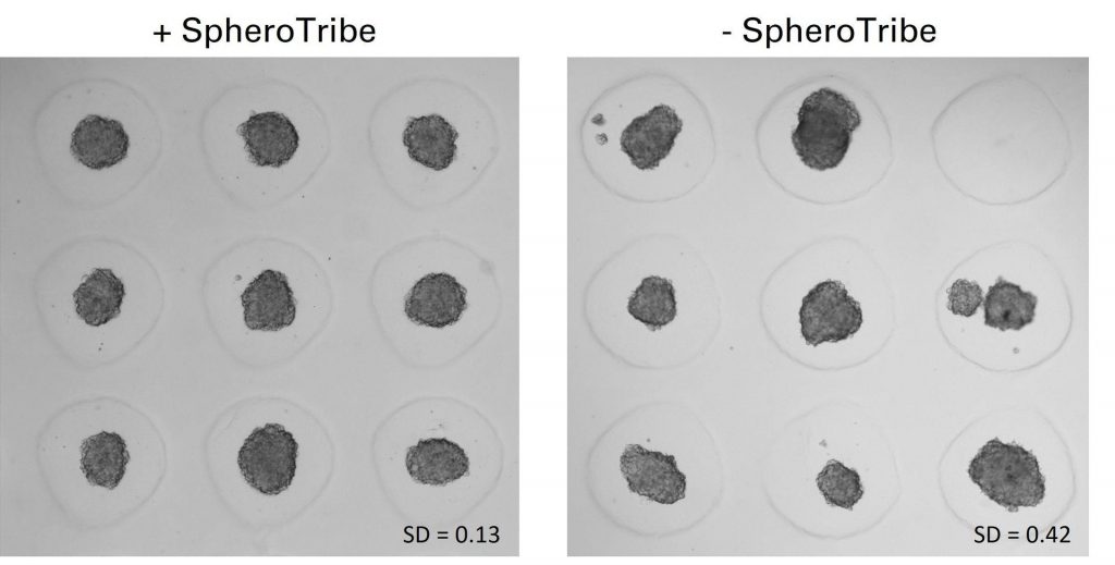
U-87 glioblastoma cells were seeded at 1,000 cells per well in round-bottom wells molded in agarose using a Stampwell U-shape (Idylle) in full culture medium without (right) or added with SpheroTribe (left). After 3 days, pictures were taken and number of cell aggregates per well were quantified from 64 independent wells and associated standard deviation (SD) values were calculated.


Kit Description:

You can purchase just the methylcellulose solution or choose among 2 different size kits:

25mL kit contents:

  • 25mL of 5X methylcellulose solution
  • 10x U-bottom 96-well plates
  • 2x racks of 96 pipette tips (200µL) with a large opening
2.5mL kit contents:

    • 2.5mL of 5X methylcellulose solution
    • 1x U-bottom 96-well plate
    • 20 pipette tips (200µL) with a large opening

Applications

SpheroTribe has been successfully used for spheroid/organoid formation with the following cell types:
Patient-derived stem-like glioblastoma cells (GB P3 and BL13), human glioblastoma cell lines (U87 & T98G), HeLa, human vaginal mucosal melanoma (HMV-II), human primary colorectal cancer cells, human breast cancer cells (MDA-MB 231), human induced pluripotent stem cells, monkey kidney fibroblast-like cell line (COS-7), primary neurons from rat embryos (E18) & murine melanoma cells (B16F10).

Experimental assays:
Once spheroids have grown to your desired size, you can use them for any kind of assay according to your regular workflow. The SpheroTribe solution can be readily washed off, leaving a spheroid available for other tests at any stage of your protocol.

Immune infiltration of B16F10 spheroids after immune checkpoint blockade

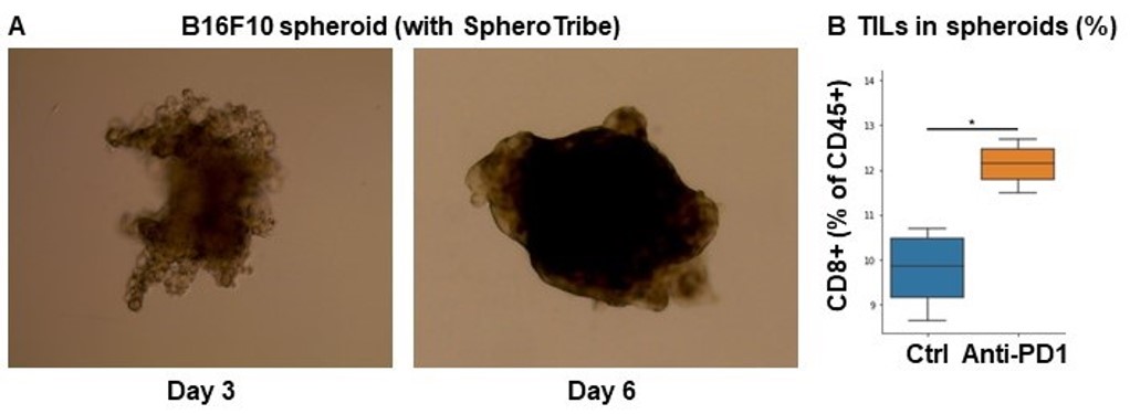
A. 10,000 B16F10 cells were grown for 6 days as spheroids using SpheroTribe. B. 100,000 PBMC from murine spleen were activated with IL-15 (40 ng/mL) [1], incubated with anti-PD1 (10 µg/mL) for 1h and added on B16F10 spheroids for 3 days.
Graph shows flow cytometry quantification of differential lymphocyte infiltration after spheroid dissociation according to treatment. N=4. Mann-Whitney U Test, p-value<0.05. [1] https://doi.org/10.3389/fonc.2022.898732.


For more information about this product or any others from the Microscopy line, contact us here.

FluoBolt- KLOTHO Fluorescence Immunoassay

High Sensitivity; Single Step Immunoassay for α-KLOTHO in Human Serum and Plasma

Eagle Biosciences is excited to highlight the FloBolt- KLOTHO Fluorescence Immunoassay from Fianostics! This assay detects only α-KLOTHO and does not cross-react with β-KLOTHO. No interference of recombinant FGF-23 with the assay’s signal up to a 100-fold molar excess was monitored. Check out the assay background and highlights below!


Background

α-KLOTHO is expressed in kidney, small intestine, placenta and prostate. The soluble peptide can be found in serum and cerebrospinal fluid. It may play a role in the calcium/ phosphorus homeostasis regulation by e.g. inhibiting active vitamin D synthesis. Further, it is also known as an antiaging-hormone by extending life span by inhibiting insulin/ IGF1 signaling pathway, as experiments in mice showed. KLOTHO is a co-receptor of fibroblast growth factor 23 (FGF-23). Research has investigated association of altered serum KLOTHO levels with chronic kidney disease and failure, renal and hepatocellular carcinomas, osteoporosis or cardiovascular diseases.

α-KLOTHO can be found either as a membrane bound or a secreted form. The membrane bound form consists of 1012 amino acids (aa), starting with a 56 aa long signaling sequence and followed by two glycosyl hydrolase 1 regions (position 57-506 and 515-953). Both glycosyl hydrolase 1 regions lack one essential Glu active site residue. Thus, it is inactive in vivo as a glycosidase although it belongs to the glycosyl hydrolase 1 family. KLOTHO’s secreted isoform, which predominates over the membrane bound form, consists of 549 amino acids (aa). It is produced by alternative splicing and differs from the membrane bound form by aa 550 to 1012 missing.


FluoBolt- KLOTHO Fluorescence Immunoassay Highlights

  • High Sensitivity
  • Single Step Procedure
  • No Washing Steps
  • No Enzyme Substrate required
  • Long Term Stable Signal

If you have any questions about this kit or any of our other offerings, contact us here.


We are excited to bring you the new Rat NT-proBNP ELISA Assay Kit! This product has been developed, validated, and manufactured by Biomedica in Austria. This assay extends our robust line of kits specific to cardiac biomarkers. Learn more below!


Biomarker Background

BNP is mainly expressed by the ventricular myocardium in response to volume overload and increased filling pressure. It is synthesized and secreted by cardiomyocytes. Mature BNP consists of 108 amino acids (proBNP or BNP-108). ProBNP is cleaved during secretion in a 1:1 ratio resulting in physiologically active BNP-32 and the biologically inactive 76 amino acid NT-proBNP. NT-proBNP (1-76) has greater plasma stability and a much longer biological half-life (90-120 minutes) than BNP, being considered as the preferred laboratory marker. BNP has a key role in cardiovascular homeostasis with biological actions including natriuresis, diuresis, vasorelaxation, and inhibition of renin and aldosterone secretion. A high concentration of BNP in the bloodstream is indicative of heart failure.


Rat NT-proBNP ELISA Assay Kit Features

  • Low Sample Volume Required – 10 µL/well
  • Controls Included
  • Sample Values Provided

Related Products

NT-proBNP ELISA Assay Kit
NT-proCNP ELISA Assay Kit
NT-proANP ELISA Assay Kit
BNP Fragment ELISA Kit


If you have any questions about this specific product or any of our other offerings, contact us here.

Fetuin A (PTM) ELISA New Product Announcement

Eagle Biosciences is excited to partner with BPM and support the Fetuin A (PTM) ELISA (DNlite-DKD)!

About Fetuin A (PTM)

The unique Fetuin A post translation modifications measured in this assay were identified in a large-scale profiling of urinary proteomics. This new biomarker can help predict the kidney condition of diabetes patients, months to years in advanced. This urine test can help predict kidney decline or complications and potentially improve a patient with diabetic kidney disease quality of care.

Principle of the Fetuin A (PTM) ELISA

The Fetuin A (PTM) ELISA (unique Fetuin-A with specific post translational modification (PTM) for Diabetic Kidney Disease (DKD)) is a competitive immunoassay. In this Fetuin A (PTM) ELISA, calibrators or unknown urine samples are mixed with anti-unique PTM Fetuin-A monoclonal antibody (mAb), and then incubated in a microplate pre-bounded with unique PTM Fetuin-A. The monoclonal antibody recognizes unique PTM Fetuin-A in calibrators or unknown samples under competition in microplate wells. After an incubation, an Horseradish Peroxide (HRP) conjugated secondary antibody is added, followed by an incubation with 3,3’,5,5’-tetramethylbenzidine (TMB) substrate. Their relative reactivity is determined by absorbance measurement at 450 nanometers (nm) and plotted by comparison with a predetermined unique PTM Fetuin-A calibration curve.

Benefits of the Assay

    • Fewer Steps
    • Shorter processing times – ever for high-throughput samples

If you have any questions about this kit or any of our other offerings, contact us here.

Cortisol ELISA Assay Kit

Eagle Biosciences is excited to announce the new Cortisol Saliva ELISA Assay Kit. The kit is intended for the quantitative determination of cortisol in human saliva.


Why Measure Cortisol?

Cortisol is the most abundant circulating steroid and the major glucocorticoid secreted by the adrenal cortex. Cortisol is physiologically effective in blood pressure maintenance and anti-inflammatory activity. It is also involved in calcium absorption, gluconeogenesis as well as the secretion of gastric acid and pepsin. It is increased under stress situations, physical exercise and external administration of ACTH. Most circulating cortisol is bound to cortisol binding globulin or transcortin and albumin. The free cortisol, which is considered the active part of blood, is about 1–2%. In the absence of appreciable amounts of the cortisol binding proteins in saliva, salivary cortisol is considered to be free and shows a diurnal rhythm with the highest levels in the morning and the lowest levels at night.

Measurement of cortisol levels in general can be used as an indicator of adrenal function and the differential diagnosis of Addison’s and Cushing’s diseases as well as adrenal hyperplasia and carcinoma.


Check out the benefits of Cortisol Saliva ELISA Assay Kit:

  • NO SHAKING required
  • ROOM TEMPERATURE incubation
  • SMALL SPECIMEN SAMPLE required

If you have any questions about this kit or our other offerings, contact us here.