Eagle Bioscience’s is excited to highlight the Alpha Synuclein Preformed Fibrils (Type 1) from StressMarq!


About the Alpha Synuclein Pre-Formed Fibrils (Type 1)

StressMarq’s Alpha Synuclein Pre-formed Fibrils (Type 1) are produced with low endotoxin levels, making them the suitable choice for both in-vivo and in-vitro work. Alpha Synuclein PFFs seed the formation of new fibrils from active alpha synuclein monomers, and can be used to induce endogenous alpha synuclein phosphorylation and subsequent Lewy body inclusion formation in neuronal cell culture or for in vitro oligomerization studies.


Background

Alpha-Synuclein (SNCA) is expressed predominantly in the brain, where it is concentrated in presynaptic nerve terminals1. Alpha-synuclein is highly expressed in the mitochondria of the olfactory bulb, hippocampus, striatum and thalamus2. Functionally, it has been shown to significantly interact with tubulin3, and may serve as a potential microtubule-associated protein. It has also been found to be essential for normal development of the cognitive functions; inactivation may lead to impaired spatial learning and working memory4. SNCA fibrillar aggregates represent the major non A-beta component of Alzheimers disease amyloid plaque, and a major component of Lewy body inclusions, and Parkinson’s disease. Parkinson’s disease (PD) is a common neurodegenerative disorder characterized by the progressive accumulation in selected neurons of protein inclusions containing alpha-synuclein and ubiquitin5, 6.

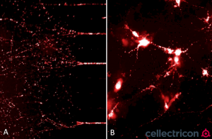
ATTO633 fluorescently-labelled alpha synuclein PFFs (SPR-322) were taken up, transported into the soma, and induced alpha synuclein aggregation in mouse neurocortical primary cells. (A) Neurites filled with fluorescently-labelled alpha synuclein seeds in a microfluidic co-culture system after 24 hours. (B) Alpha synuclein seeds within the soma and neurites of mouse neurocortical primary cells after 24 hours. Experiment and imaging courtesy of Cellectricon.

References

  1. “Genetics Home Reference: SNCA”. US National Library of Medicine. (2013).
  2. Zhang L., et al. (2008) Brain Res. 1244: 40-52.
  3. Alim M.A., et al. (2002) J Biol Chem. 277(3): 2112-2117.
  4. Kokhan V.S., Afanasyeva M.A., Van’kin G. (2012) Behav. Brain. Res. 231(1): 226-230.
  5. Spillantini M.G., et al. (1997) Nature. 388(6645): 839-840.
  6. Mezey E., et al. (1998) Nat Med. 4(7): 755-757.

The Alpha Synuclein Preformed Fibrils (Type 1) were utilized in the following studies:


If you have any questions about this product or any of our other offerings, contact us here.

GA-Map Validated on MAGPIX

The Genetic Analysis GA-Map® Dysbiosis Text Lx has been validated on the Luminex MAGPIX system. The company proudly announced the successful completion of validation at the beginning of February. Previously, the GA-Map Dysbiosis Test Lx was only validated for the Luminex 200.

GA-Map Dysbiosis Test Lx Principle

The GA-Map Dysbiosis Test Lx is a tool that allows mapping of the intestinal microbiota profile for a selected set of bacteria in a sample. The GA-Map Dysbiosis Test is based on advances in DNA profiling using probes targeting variable regions (V3 to V7) of the bacterial 16S rRNA gene to characterize and identify bacteria present. The targets are identified in a molecular multiplex assay that utilizes the Single Nucleotide Primer Extension (SNuPE) technology patented by professor Knut Rudi of the Norwegian University of Life Sciences (​NMBU) (US6617138). A unique algorithm takes advantage of all the data generated by the detection of the SnuPE products to determine the microbiota profile in the sample. The algorithm is incorporated in the GA-Map Dysbiosis Analyzer software that accompanies the test.

Luminex MAGPIX Validation

The MAGPIX® instrument is the most affordable instrument of the Luminex’s xMAP instruments. The MAGPIX system is already a widely used platform and a large number of laboratories have already installed this system for performing other tests.

The human microbiome market is accelerating both in terms of evidence-based research and pharma products launched. The market’s need for a validated microbiome test is growing with this market. Thus, the news today that more labs can start microbiome testing with GA-Map on MAGPIX will fuel this growth and create more research opportunities.

Check out the full press release here.


If you have any questions about the GA-Map Dysbiosis Test Lx or our other offerings, contact us here.


New data suggests the Cov19 FluoBolt-DAT detects antibodies against the new Omicron variant.

Quantitative Duplex Antibody Test (DAT) for antibodies to SARS-CoV-2

The Cov19 FluoBolt-DAT allows the simultaneous and quantitative measurement of antibodies against the S1 RBD AND Nucleocapsid antigen of SARS-CoV-2. In contrast, other antibody assays detect all antibodies generated against the S1- OR NC-protein. This test, however, is epitope specific. It detects immune-dominant antibodies species, i.e. the most important antibodies generated by either vaccination or infection.

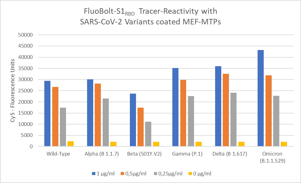
This is demonstrated by the fact, that it detects anti-S1RBD against all dominant virus variants identified so far. The data indicates (see chart below) that the S1RBD part of the Cov19 FluoBolt-DAT assay detects the immune-dominant epitope and that individuals testing S1RBD -positive in the FluoBolt-DAT test have developed significant amounts of antibodies against this epitope. Therefore, they might be well protected against all current and future SARS-CoV-2 variants

Cov19 FluoBolt-DAT

Benefits of the Cov19 FluoBolt-Dat Assay:

    • Only One Single Measurement Required
    • Small Sample Size Need – 10 uL
    • Results within 60 minutes

    Other Cov19 FluoBolt-DAT Resources:


    Check out our other COVID-19 assays here.

    If you have any questions about this kit or our other offerings, contact us here.

GA-map Technical Spotlight

This week Eagle Biosciences is highlighting a revolutionary test in the microbiome industry. The GA-map® Dysbiosis Test Lx was developed by Genetic Analysis in Norway. The GA-map ® is a tool that allows mapping of the intestinal microbiota profile for a selected set of bacteria in a sample. The test is based on advances in DNA profiling using probes targeting variable regions (V3 to V7) of the bacterial 16S rRNA gene to characterize and identify bacteria present. The targets are identified in a molecular multiplex assay that utilizes the Single Nucleotide Primer Extension (SNuPE) technology patented by professor Knut Rudi of the Norwegian University of Life Sciences (​NMBU) (US6617138). A unique algorithm takes advantage of all the data generated by the detection of the SnuPE products to determine the microbiota profile in the sample. The algorithm is incorporated in the GA-map® Dysbiosis Analyzer software that accompanies the test.

Check out this video for a quick overview of the GA-map ® Dysbiosis Test Lx Platform equipment and techniques.

Note: This test is for research use only and not to be used for diagnostic procedures


If you have any questions about the GA-map® Dysbiosis Test Lx or our other offerings contact us here.


Polystreptavidin R (Polystrept R) reagent is for the surface coating of plastics, membranes, beads and etc. with maximum biotin binding capacity. It improves the signal-noise-relation in testing and can help save on material costs.

About Polystreptavidin R (Polystrept R)

Polystrept R is a chemically modified polymerized Streptavidin characterized by an extraordinary high Biotin binding capacity. Polystrept R coatings on solid phases offer a universal immobilization principle for the detection and analysis of proteins, peptides, PCR-fragments, haptens etc., which must be present in a biotinylated form. Coatings made of Polystreptavidin R combine the excellent binding capacity with a high chemo and thermo stability and a long shelf life. It is suitable for coatings of membranes, beads, biochips, plastics etc.

Learn about the benefits of using Polystrept R in your research in this video from Biotez!


Why use streptavidin-coated microplates and Polystrept R with lateral flow test strips??

  • Convenient method for a uniform and stable immobilization of biotinylated molecules
  • Immobilization of Biomolecules on solid phases in a directed manner, while avoiding or minimizing denaturation
  • Ready-to-use, no washing or blocking steps required
  • High signal-to-noise ratio
  • Low nonspecific binding
  • Long shelf life
  • Lower reagent consumption and especially enormous cost savings with lateral flow test stripes

Related Posts


If you have any questions about Polystreptavidin R or our other offerings, contact us here.

ELISA for Detecting SARS-CoV-2 Mutant Strains

SARS-CoV-2, a single stranded RNA virus, has quickly become the subject of much research worldwide. Coronaviruses are composed of four primary proteins: the spike (S), envelope (E), membrane (M), and nucleocapsid (N). The spike protein (S-protein) performs two primary tasks that aid in host infection: 1) mediates the attachment between the virus and host cell surface receptors such as ACE2, and 2) facilitates viral entry into the host cell by assisting in the fusion of the viral and host cell membranes. In December of 2020 three new SARS-CoV-2 variants came to light, UK B.1.1.7, South Africa B.1.351 and Brazil P.1/P.2.

Eagle Biosciences offers a SARS CoV-2 Spike Protein Antigen Quantitative ELISA Assay Kit that is highly sensitive. Clinical studies show that this kit is effective at detecting the spike protein antigen in all three major circulating SARS-CoV-2 variant strains. Click here to learn more.

About the SARS-CoV-2 Spike Protein Antigen Quantitative ELISA Assay Kit

  • Validated for both serum and plasma samples.
  • No cross-reactivity with other respiratory pathogens.
  • Standardization and reproducibility, with lot-to-lot consistency.

Eagle Biosciences is dedicated to providing support to researchers in their studies of SAR-CoV-2 and the new variants. To hear more about our COVID-19 resources and other offerings, contact us here.

iLite Assay Ready Cells

iLite® Assay Ready Cells are developed by our partners at Svar Life Science. iLite technology is based upon a reporter gene assay format, modified and adapted for applications during the whole drug development cycle as well as for monitoring of biological drugs. These cell lines can be developed for any biopharmaceutical target and assays for drug potency, i.e. drug activity, and neutralizing antibodies (NAbs) can easily be set-up using the same cell line. The Assay Ready cells are genetically engineered to be used with a reporter gene assay technique for detection the the drug potency and the NAbs.


A Message from SVAR about iLite:

“Many therapeutic antibodies that are used to treat common disorders, such as cancer and autoimmune diseases work – at least in part – by activating ADCC.

Our reporter gene-based iLite® ADCC bioassays offer a convenient and powerful way of measuring the ability of an antibody to elicit ADCC.

Read our new white paper and learn about the performance of our ADCC bioassays and how they compare to a leading competitor in terms of sensitivity and dynamic range.”

Download the White Paper here.


Find all of the iLite products offered on the iLite Assay Ready Cells product page.

ELISpot Kits

Eagle Biosciences proudly offers a broad range of products in our extensive catalog. In addition to the well known ELISA format, we offer several assay kits in a enzyme-linked immunospot (ELISpot) assay format.

ELISA vs. ELISpot

ELISAs use antibodies to detect the presence and concentration of a protein in a liquid sample. In the most common form, antigens are applied to a stable surface (usually a microtiter plate). Then the corresponding antibody, linked with an enzyme, is added to form a complex. Finally, the enzyme’s substrate is added, which produces a measurable reaction, usually in the form of a color change.

ELISpots also utilize antibody-enzyme reactions. However, instead of measuring the concentration of protein in a sample, they measure the number of cells secreting the cytokine of interest in a sample. Antigens are applied to the plate surface, and when samples are added, cells settle on the bottom of the wells. The cytokines secreted by the cells are captured by the surrounding antigens. After the appropriate incubation time, the cells are removed and the secreted cytokine is detected using a detection antibody linked with an enzyme. The enzyme’s substrate is added, producing a reaction. The end result is visible spots on the on the surface of the plate. Each spot corresponds to an individual cell that was secreting the target protein.

The information provided by ELISpot kits is applicable to various fields of study including: transplantation, vaccine development, T-cell regulation analysis, autoimmune disease, cancer, allergy, and viral infections. These kits are highly sensitive and easy to use.

ELISpot Kit Procedure

Our available kits:

IFN gamma ELISpot Assay Kit

Mouse IFN gamma ELISpot Assay Kit

Rat IFN gamma ELISpot Assay Kit

TNF alpha ELISpot Assay Kit

Rat TNF alpha ELISpot Assay Kit

Contact us if you are looking for a specific ELISpot or if you have questions about any of our products.

A new article published by Nelly Kanberg, et al. titled Neurochemical evidence of astrocytic and neuronal injury commonly found in COVID-19 has set out to study how COVID-19 impacts the central nervous system. It’s already known how this novel coronavirus effects the respiratory system, but does the body’s inflammatory response to this virus cause lasting effects to the nervous system?

For this study, two biomarkers in human plasma were measured; neurofilament light chain protein (NfL) and glial fibrillary acidic protein (GFAP). The samples came from patients who had mild, moderate or severe cases of COVID-19 (n=47). Of those 47 patients, the ones who had a severe case of COVID-19 showed higher concentrations of GFAP and NfL.

Glial Fibrillary Acidic Protien, or GFAP for short, is a known biomarker in the central nervous system (CNS) that is typically studied in those with brain injuries. When a brain injury occurs (concussion, retinal stess, tumors, etc.), GFAP is released into the blood stream and helps determine the severity of the injury. The results of this study help support and determine the relationship between COVID-19 and potentially lasting neural injuries.

To read this article in full, click here.

To learn more about how to measure GFAP, click here or contact us with your questions

Eagle Biosciences proudly offers a broad range of products in our extensive catalog. In addition to the well known ELISA format, we offer several assay kits in a Chemiluminescent Enzyme Immunoassay (CLEIA) format.

ELISA vs. CLEIA

ELISAs use antibodies to detect the presence of a protein in a liquid sample. In the most common form, antigens are applied to a stable surface (usually a microtiter plate). Then the corresponding antibody, linked with an enzyme, is added to form a complex. Finally, the enzyme’s substrate is added, which produces a measurable reaction, usually in the form of a color change.

CLEIAs also utilize antibody-enzyme reactions, but offer a higher level of sensitivity due use of chemiluminescence rather than colorimetric absorbance or fluorescence. Chemiluminescence allows detection of trace amounts of analytes because samples typically have little to no background interference. These reactions are also easily measured using a photomultiplier or photodiode. These types of kits are increasing in popularity due to their increased sensitivity, specificity, reliability, and reproducibility.

Our available CLEIA/CLIA kits

GFAP (Glial Fibrillary Acidic Protein) CLIA Assay Kit
MedFrontier FGF23 (Human Intact FGF23) CLEIA ELISA Assay

Contact us if you are looking for a specific CLEIA or if you have questions about any of our products.