FluoBolt Asporin Fluorescence Immunoassay

$1,350.00

The FluoBolt™-Asporin Fluorescence Immunoassay is intended for the quantitative determination of asporin in human serum. The FluoBolt™-Asporin Fluorescence Immunoassay has been linked to conditions like osteoarthritis, lumbar disc degeneration and tumor progression research.

SKU: FIA-1702 Category: Tags: ,

FluoBolt Asporin Fluorescence Immunoassay

The FluoBolt Asporin Fluorescence Immunoassay is manufactured in Austria by Fianostics GmbH

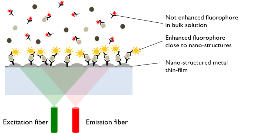
Method: Metal Enhanced Direct Sandwich Fluorescence Immunoassay
Size: 1×96 wells
Sensitivity: LOD <0 pmol/l + 3 SD): 10 pmol/l; LLOQ: 25 pmol/l
Dynamic Range: 0-200 pmol/l
Incubation Time: Overnight
Sample Type: Serum
Sample Size: 10 µl
For Research Use Only

This platform is fully compatible to standard laboratory methodology using 96 well microtiter plate format and assays based on this technology can be run on any standard fluorescence microplate reader.


This assay is regularly delivered standard with AlexaFluor680 labeled detection-antibody. If you want to use a different dye, the following options are available:

  • FITC labeled (FIA-1702-F)
  • Cy3 labeled (FIA-1702-C3)
  • Cy5 labeled (FIA-1702-C5)

Conversion factor: 1 pg/ml = 0.018 pmol/l (MW: 55,7 kD / Monomer)


Assay Background

Asporin, also known as periodontal ligament-associated protein 1 (PLAP1) is a dimeric secreted extracellular matrix protein, which belongs to the small leucine-rich proteoglycan (SLRP) family. It consist out of 380 amino acids and has a highly conserved pro peptide sequence which contains a series of leucine rich repeats and are flanked by two cysteine residues in the C Terminal region. Further it has four cysteine residues that form disulphide bonds as well as aspartic acid repeats in the N-Terminal region.

High levels of Asporin can be found in aorta, uterus and osteoarthritic articular cartilage. Further, moderate levels of aspirin expression can be found in small intestine, heart liver, bladder, ovary, stomach, the adrenal-, thyroid-, and mammary gland. Lower levels of expression can be seen in the trachea, bone marrow and lung. Asporin is known to negatively regulate PDL differentiation and mineralisation as well as it inhibits BMP- dependent activation of SMAD proteins. Further, it directly binds to TGFb-1 subsequently, binds to collagen by way of its LRR domain. Through its interaction with TGFb-1, Asporin negatively regulates chondrogenesis in the articular cartilage by blocking the TGF-beta/receptor interaction on the cell surface and inhibiting canonical TGF-beta/Smad signalling. Moreover, it has the ability to bind calcium, giving it regulatory properties in osteoblast driven mineralisation and regulates FGF2 through direct and indirect interactions. Next to its regulatory properties in terms of cartilage and bone homeostasis, Asporin expression has also been linked to cancer invasion and progression. However, its value as biomarker remains to be established yet.


ASPORIN has been linked to:

  • Osteoarthritis
  • Lumbar Disc Degeneration
  • Tumor Progression

Related Products

FluoBolt-Klotho Fluorescence Immunoassay
FluoBolt-Periostin Fluorescence Immunoassay
FluoBolt Noggin Fluorescence Immunoassay

Additional Information

About Metal Enhanced Fluorescence


Metal Enhanced Fluorescence (MEF) using metal nano-structures offers the possibility to increase the analytical sensitivity of systems based on Iuorescence detection dramatically. FIANOSTICS has developed a new MEF- immunoassay platform, that allows up to 300 fold gains of sensitivity. This platform is fully compatible to standard laboratory methodology using 96 well microtiter plate format and assays based on this technology can be run on any standard fIuorescence microplate reader. Its unique features enable us to develop high sensitivity, single step with no wash florescence immunoassays for low abundance biomarkers.
MEF Principle

Assay Principle


All reagents must be at room temperature (18-26C) before use in this assay

  • Add 50 μl of the selected fluorescence labeled detection antibody (EAF or EA3 or EA5 or EAA) to all wells required. Swirl gently.
  • Add 10 μl of standard, control or sample to the wells according to the marked positions on the protocol sheet, swirl gently, cover tightly with the delivered adhesive film and incubate over night at room temperature (18-26°C) in the dark.
  • a)If your reader allows bottom reading, read the plate without any further processing at the Ex/Em wavelength fitting to the delivered detection antibody (495/518 nm for EAF, 550/570 nm for EA3, 650/670 nm for EA5, 679/702 nm for EAA). Gain should be set to achieve at least 10000 Iuorescence units (F.U.) between the signal of the 0 pM and the 200 pM ASPORIN standard. Samples with signals exceeding the signal of the highest standard must be re-run with an appropriate dilution using sample diluent (ED).
    b)If your reader has no bottom read option or if you want to store the plate for documentation purposes, discard or aspirate the content of the wells and wash 3x with diluted wash buffer. Use a minimum of 200 μl wash buffer per well. After the final wash, remove remaining fIuid by strongly tapping the plate against a paper towel. Read the plate in top con_guration without any further processing at the Ex/Em wavelength fitting to the chosen detection antibody (495/518 nm for EAF, 550/570 nm for EA3, 650/670 nm for EA5, 679/702 nm for EAA). Gain should be set to achieve at least 10000 Iuorescence units (F.U.) between the signals of the 0 pM and the 200 pM ASPORIN standard. Samples with signals exceeding the signal of the highest standard must be re-run with appropriate dilution using sample diluent
    Quality of bottom reading (3a) may vary between microplate readers. For first time users we suggest to perform the washing step and follow protocol 3b (ED).
  • Store the plate with desiccant at 4°C (2-8°C) in the aluminium bag. Unused wells are stable until expiry date stated on the label. Fluorescence signals of standards, controls and samples remain detectable for at least two month at the plate surface, depending on signal intensity achieved.

Package Inserts


Product Citations