FluoBolt Periostin Fluorescence Immunoassay

$1,465.00

The FluoBolt™-Periostin Fluorescence Immunoassay is intended for the quantitative determination of periostin in human serum. The FluoBolt™-Periostin Fluorescence Immunoassay has been linked to various progression and matastasis associated with pancreatic, ovarian, lung, breast, colon, gastric, thyroid and esophageal tumors.

SKU: FIA-1703 Category: Tags: ,

FluoBolt Periostin Fluorescence Immunoassay

The FluoBolt Periostin Fluorescence Immunoassay is manufactured in Austria by Fianostics GmbH

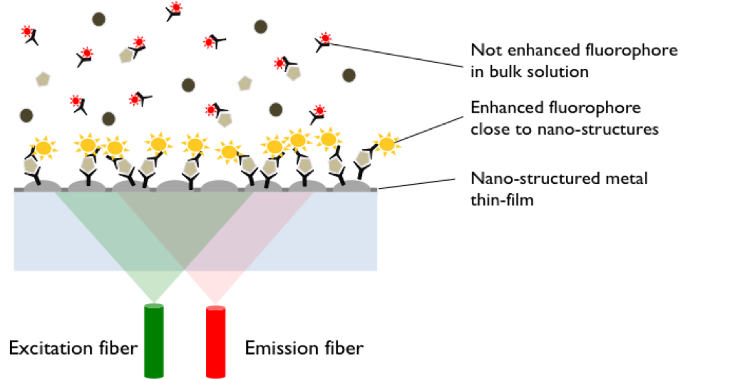
Method: Metal Enhanced Direct Sandwich Fluorescence Immunoassay
Size: 1×96 wells
Sensitivity: LOD <0 pmol/l + 3 SD): 2 pmol/l; LLOQ: 11 pmol/l
Dynamic Range: 0-180 pmol/l
Incubation Time: Overnight
Sample Type: Serum
Sample Size: 20 µl
For Research Use Only

This platform is fully compatible to standard laboratory methodology using 96 well microtiter plate format and assays based on this technology can be run on any standard fluorescence microplate reader.


This FluoBolt-Periostin Fluorescence Immunoassay is regularly delivered standard with AlexaFluor680 labeled detection-antibody. If you wish to use a different dye, the following options are available:

  • FITC labeled (FIA-1703-F)
  • Cy3 labeled (FIA-1703-C3)
  • Cy5 labeled (FIA-1703-C5)

Conversion factor: 1 ng/ml = 11 pmol/l (MW: 93.3 kD)


Assay Background

PERIOSTIN (UniProtKB – Q15063), also known as osteoblast specific factor 2 (OSF-2), is a cell adhesion protein belonging to the fasciclin domain-containing protein family. It consists of 836 amino acids (aa) starting with a 21 aa long signalling sequence, followed by a Emilin-like domain rich in cysteine, four repeated fasiclin 1 and a C-terminal variable domain, which is different among the 7 splice variants (isoforms) in humans.

PERIOSTIN is expressed during ontogenesis as well as in adult connective tissues submitted to mechanical stress such as bone, tendons, heart valves, skin and the periodontal ligaments. Further, it is expressed in aorta, stomach, lower gastrointestinal tract, placenta, uterus, thyroid and breast tissue. In bone, PERIOSTIN directly interacts with collagen type I, fibronectin, Notch 1, tenascin-C and BMP-1, resulting in enhanced proteolytic activation of lysyl oxidase for collagen cross-linking stabilising bone matrix. Next to developing, maintaining and repairing tissue, PERIOSTIN plays a vital role in tumorigenesis by interacting with various cell-surface receptors and signaling pathway, which e.g. results in inactivation of integrin- mediated signaling, leading to promoting cell adhesion and motility which is of relevance for tumor progression and metastasis. Elevated serum PERIOSTIN levels have been associated with pancreatic, ovarian, lung, breast, colon, gastric, thyroid and oesophageal tumours.


PERIOSTIN has been associated with the following tumors:

  • Pancreatic
  • Ovarian
  • Lung
  • Breast
  • Colon
  • Gastric
  • Thyroid
  • Esophageal

Related Products

FluoBolt-Klotho Fluorescence Immunoassay
FluoBolt Noggin Fluorescence Immunoassay
FluoBolt Asporin Fluorescence Immunoassay

Additional Information

About Metal Enhanced Fluorescence


Metal Enhanced Fluorescence (MEF) using metal nano-structures offers the possibility to increase the analytical sensitivity of systems based on Iuorescence detection dramatically. FIANOSTICS has developed a new MEF- immunoassay platform, that allows up to 300 fold gains of sensitivity. This platform is fully compatible to standard laboratory methodology using 96 well microtiter plate format and assays based on this technology can be run on any standard fIuorescence microplate reader. Its unique features enable us to develop high sensitivity, single step with no wash florescence immunoassays for low abundance biomarkers.

Assay Principle


All reagents must be at room temperature (18-26C) before use in this assay

  • 1) Add 40 µl of the selected fluorescence labeled detection antibody (FAF or FA3 or FA5 or FAA) to all wells required. Swirl gently.
  • Add 20 μl of standard, control or sample to the wells according to the marked positions on the protocol sheet, swirl gently, cover tightly with the delivered adhesive film and incubate over night at room temperature (18-26°C) in the dark.
  • a)If your reader allows bottom reading, read the plate without any further processing at the Ex/Em wavelength fitting to the delivered detection antibody (495/518 nm for FAF, 550/570 nm for FA3, 650/670 nm for FA5, 679/702 nm for FAA). Gain should be set to achieve at least 10000 fluorescence units (F.U.) between the signal of the 0 pM and the 180 pM PERIOSTIN standard. Samples with signals exceeding the signal of the highest standard must be re-run with an appropriate dilution using sample diluent (FD).
    b)If your reader has no bottom read option or if you want to store the plate for documentation purposes, discard or aspirate the content of the wells and wash 3x with diluted wash buffer.Use a minimum of 200 µl wash buffer per well. After the final wash, remove remaining fluid by strongly tapping the plate against a paper towel. Read the plate in top configuration without any further processing at the Ex/Em wavelength fitting to the chosen detection antibody (495/518 nm for FAF, 550/570 nm for FA3, 650/670 nm for FA5, 679/702 nm for FAA).
    Quality of bottom reading (3a) may vary between microplate readers. For first time users we suggest to perform the washing step and follow protocol 3b (ED).
  • Store the plate with the 2 desiccant bags supplied at 4°C (2-8°C) in the aluminum bag. Unused wells are stable until expiry date stated on the label. Fluorescence signals of standards, controls and samples remain detectable for at least two month at the plate surface, depending on signal intensity achieved.

Package Inserts