Our Homoarginine ELISA Kit was highlighted in a recent publication! Researchers compiled what is known of homoarginine’s pathways and its role in different diseases and conditions. Check out the abstract and access to the full text below!


Abstract

Purpose: Homoarginine (hArg) is an arginine metabolite that has been known for years, but its physiological role in the body remains poorly understood. For instance, it is well known that high hArg concentrations in the blood are protective against several disease states, yet the mechanisms behind these health benefits are unclear. This review compiles what is known about hArg, namely its synthetic pathways, its role in different diseases and conditions, and its proposed mechanisms of action in humans and experimental animals.

Findings: Previous work has identified multiple pathways that control hArg synthesis and degradation in the body. Furthermore, endogenous hArg can modulate the cardiovascular system, with decreased hArg being associated with cardiovascular complications and increased mortality. Studies also suggest that hArg could serve as a diagnostic biomarker for a variety of immune, pancreatic, renal, and hepatic dysfunctions. Finally, in women, hArg concentrations rapidly increase throughout pregnancy and there are suggestions that alterations in hArg could indicate pregnancy complications like pre-eclampsia.

Summary: Homoarginine is an under-appreciated amino acid with potential wide-ranging roles in systemic health, pregnancy, and pathophysiology. Although recent research has focused on its health or disease associations, there is a need for more investigations into understanding the mechanistic pathways by which hArg may operate. This could be aided using metabolomics, which provides a comprehensive approach to correlating multiple metabolites and metabolic pathways with physiological effects. Increasing our knowledge of hArg’s roles in the body could pave the way for its routine use as both a diagnostic and therapeutic molecule

Zubkowski A, Sferruzzi-Perri AN, Wishart DS. Mechanisms of Homoarginine: Looking Beyond Clinical Outcomes. Acta Physiol. 2025; 241:e14273. doi:10.1111/apha.14273


If you have any questions about this product or any of our offerings, contact us here.

The MMAE Antibody Drug Conjugate (ADC) ELISA Assay Kit was referenced in a recent publication! Scientists aimed to develop an antibody-drug conjugate (ADDC) to act against MET and RON receptors to treat cancers with high phenotypic heterogeneity. View the abstract and access to the full text below!


Abstract

Cancer heterogeneity, characterized by diverse populations of tumorigenic cells, involves the occurrence of differential phenotypes with variable expressions of receptor tyrosine kinases. Aberrant expressions of mesenchymal–epithelial transition (MET) and recepteur d’origine nantais (RON) receptors contribute to the phenotypic heterogeneity of cancer cells, which poses a major therapeutic challenge. This study aims to develop a dual-targeting antibody–drug conjugate (ADC) that can act against both MET and RON for treating cancers with high phenotypic heterogeneity. Through immunohistochemical staining, we show that MET and RON expressions are highly heterogeneous with differential combinations in more than 40% of pancreatic and triple-negative breast cancer cases. This expressional heterogeneity provides the rationale to target both receptors for cancer therapy. A humanized bispecific monoclonal antibody specific to both MET and RON (PCMbs–MR) is generated through IgG recombination using monoclonal antibody sequences specific to MET and RON, respectively. Monomethyl auristatin E is conjugated to PCMbs–MR to generate a dual-targeting ADC (PCMdt–MMAE), with a drug-to-antibody ratio of 4:1. Various cancer cell lines were used to determine PCMdt-MMAE-mediated biological activities. The efficacy of PCMdt–MMAE in vivo is evaluated using multiple xenograft tumor models. PCMdt–MMAE shows a favorable pharmacokinetic profile, with a maximum tolerated dose of ~30 mg/kg in mice. Toxicological studies using Sprague–Dawley rats reveal that PCMdt–MMAE is relatively safe with slight-to-moderate, temporary, and reversible adverse events. Functionally, PCMdt-MMAE induces a robust internalization of both MET and RON and causes a large-scale cell death in cancer cell lines exhibiting MET and RON heterogeneous co-expressions. Both in vitro and in vivo studies demonstrate that the dual-targeting approach in the form of an ADC is highly effective with a long-lasting effect against tumors exhibiting MET/RON heterogeneous phenotypes. Hence, we can suggest that a dual-targeting ADC specific to both MET and RON can be employed as a novel therapeutic strategy for tumors with expressional phenotypic heterogeneity.

Wang, M., Ma, Q., Suthe, S.R. et al. Humanized dual-targeting antibody–drug conjugates specific to MET and RON receptors as a pharmaceutical strategy for the treatment of cancers exhibiting phenotypic heterogeneity. Acta Pharmacol Sin 46, 1375–1389 (2025). https://doi.org/10.1038/s41401-024-01458-7


If you have any questions about this product or any of our other offerings, contact us here.

The Calprotectin ELISA Assay Kit was utilized in a recent publication! Researchers investigated whether cytotoxic T-lymphocyte-associated protein 4 deficiency (CTLA4-D), a rare genetic disorder, could be distinguished from an individuals’ microbiome. They also aimed to identify biomarkers to indicate disease severity. Check out the abstract and access to the full text below!


Abstract

Background: Cytotoxic T-lymphocyte-associated protein 4 deficiency (CTLA4-D) is an inborn error of immunity (IEI) caused by heterozygous mutations, and characterized by immune cell infiltration into the gut and other organs, leading to intestinal disease, immune dysregulation and autoimmunity. While regulatory T-cell dysfunction remains central to CTLA4-D immunopathogenesis, mechanisms driving disease severity and intestinal pathology are unknown but likely involve intestinal dysbiosis. We determined whether the intestinal microbiome and metabolome could distinguish individuals with severe CTLA4-D and identify biomarkers of disease severity.

Results: The genera Veillonella and Streptococcus emerged as biomarkers that distinguished CTLA4-D from healthy cohorts from both the National Institutes of Health (NIH) Clinical Center, USA (NIH; CTLA-D, n = 32; healthy controls, n = 16), and a geographically distinct cohort from the Center for Chronic Immunodeficiency (CCI) of the Medical Center – University of Freiburg, Germany (CCI; CTLA4-D, n = 25; healthy controls, n = 24). Since IEIs in general may be associated with perturbations of the microbiota, a disease control cohort of individuals with common variable immunodeficiency (CVID, n = 20) was included to evaluate for a CTLA4-D-specific microbial signature. Despite common IEI-associated microbiome changes, the two bacterial genera retained their specificity as biomarkers for CTLA4-D. We further identified intestinal microbiome and metabolomic signatures that distinguished patients with CTLA4-D having severe vs. mild disease. Microbiome changes were associated with distinct stool metabolomic profiles and predicted changes in metabolic pathways. These differences were impacted by the presence of gastrointestinal manifestations and were partially reversed by treatment with abatacept and/or sirolimus.

Conclusions: Loss of intestinal microbial diversity and dysbiosis causing metabolomic changes was observed in CTLA4-D. Albeit some of these features were shared with CVID, the distinct changes associated with CTLA4-D highlight the fact that IEI-associated microbiome changes likely reflect the underlying immune dysregulation. Identified candidate intestinal microbial and metabolic biomarkers distinguishing individuals with CTLA4-D based on severity should be studied prospectively to determine their predictive value, and investigated as potential therapeutic targets.

Chandrasekaran P, et al. The intestinal microbiome and metabolome discern disease severity in cytotoxic T-lymphocyte-associated protein 4 deficiency. Microbiome. 2025 Feb 11;13(1):51. doi: 10.1186/s40168-025-02028-7. Erratum in: Microbiome. 2025 Mar 15;13(1):74. doi: 10.1186/s40168-025-02069-y. PMID: 39934899; PMCID: PMC11817180.


If you have any questions about this product of any of our other offerings, contact us here.

The Anti-CCP ELISA Assay Kit played a key role in a new publication! This study aimed to understand pathways leading to B cell activation and autoantibody production, and identify new biomarkers that could be helpful in predicting and assessing the response to abatacept in rheumatoid arthritis patients who don’t respond well to methotrexate and other standard treatments. Dive into the abstract and full paper below!


Abstract

Objectives: To investigate whether biomarkers related to B cell activation and autoantibody production are associated with the response to abatacept in rheumatoid arthritis (RA) patients.

Methods: Twenty-five patients with RA were enrolled in this study. Responders (n=10) to abatacept were subjects who achieved ACR50 response at week 24. Serum levels of soluble biomarkers were measured with ProcartaPlex by Luminex or ELISA. Peripheral blood mononuclear cells were isolated and analysed for T cell and B cell subsets by flow cytometry. Patients were genotyped for human leukocyte antigen (HLA)-DRB1 shared epitope (SE) alleles. Baseline levels and longitudinal changes of markers were assessed between responders and nonresponders.

Results: Baseline levels of anti-cyclic citrullinated peptide (anti-CCP) antibodies (p=0.01), IgM rheumatoid factor (RF) (p=0.02), CXC chemokine ligand 13 (CXCL13, p=0.02), sCD23 (p<0.05), as well as frequencies of CD19+CD11c+IgD-CD27- B cells (p=0.04), were higher in responders than nonresponders. Among them, anti-CCP and frequencies of CD19+CD11c+IgD-CD27- B cells were independently associated with response to abatacept. The presence of two alleles of SE was associated with responders (p=0.04). Patients with 2 alleles of SE had higher levels of anti-CCP (p=0.02) and IgM RF (p=0.04) compared to patients with 0 or 1 allele. Further, IgM RF and CXCL13 levels decreased only in responders (p=0.02 and 0.004 respectively, at week 24), while anti-CCP levels did not decrease significantly in either responders or nonresponders.

Conclusion: Markers of B cell activation including anti-CCP and frequencies of CD19+CD11c+IgD-CD27- B cells in RA were associated with response to abatacept. IgM RF and CXCL13 decreased only in responders and could be potentially used as pharmacodynamic markers.

Wang T, Giltiay NV, Lood C, Wang N and Han BK (2025) Evaluation of B cell related markers and autoantibodies in rheumatoid arthritis patients treated with abatacept. Front. Immunol. 16:1504454. doi: 10.3389/fimmu.2025.1504454


If you have any questions about this product or any of our other offerings, contact us here.

Eagle Biosciences is excited to highlight our product series for Host Cell Protein Detection!

Host cell proteins (HCPs) are a major class of impurities produced during biotherapeutic manufacturing. They must be removed from the final drug product to both assure patient safety and maintain drug efficacy. Our wide range of Host Cell Protein Detection Kits are easy to use and highly sensitive.


What are HCPs and why must they be removed from biologic drugs?

HCPs are proteins produced or encoded by the host organisms used to produce recombinant therapeutic proteins. Genetic engineering allows the host organism cells to be transformed to produce a protein of interest. During the recombinant protein production, host cells also coproduce proteins related to the normal cell functions such structural proteins, as well as proteins required for normal cellular growth and function, and vary in both number and concentration depending on the chosen host species and the manufacturing process being used. In general, apart from the therapeutic protein of interest, all endogenous proteins co-expressed by the host cells are called host-cell proteins.

Why must HCPs be removed from biologic drugs?

HCPs must be removed from the final biotherapeutic product to avoid adverse effects. Almost all HCPs carry safety risks as foreign proteins due to the potential to elicit immune response in humans (e.g, cytokine storm). In addition, some HCPs can also act to enhance the immune response to a drug product. Certain HCPs can also affect drug product stability and efficacy if not adequately removed or inactivated.

How are HCPs detected?

ELISAs are widely used for detecting HCPs, where they are generally configured in a sandwich assay format for improved specificity. In this scenario, a microplate-bound antibody is used for analyte capture, then a second analyte-specific antibody (that binds a different epitope on the target molecule) is added to enable detection. By incorporating a reference standard (e.g., a purified protein) into the assay design, it is possible to quantify the analyte of interest and confirm that its concentration meets regulatory requirements. Advantages of ELISA are that it is sensitive and compatible with high sample throughput – key considerations for biopharmaceutical manufacturing.


If you have any questions about these products or any of our other offerings, contact us here.

iLite Assay Ready Cells

iLite® Assay Ready Cells are developed by our partners at Svar Life Science. The cell based solutions are based on the iLite technology, a cleverly engineered cell-based assay system with a dual reporter gene readout. They offer the ease of use and robustness of a Ligand Binding Assay and can be developed for virtually any pharmaceutical target and allows an easy, rapid and accurate test format for measurement and quantification of drug activity and immunogenicity.


Key Benefits

Assay Ready Format:
iLite cells are delivered as Assay Ready Cells, and stored at -80°C. There is no need for cell culturing and continuous maintenance of cells – just thaw and dilute before use in the assay.

Normalization Gene:
A second reporter gene used as an internal control will compensate for differences in cell number. In addition, it can be used to compensate for serum matrix effects, other complex matrices or if luciferase is sensitive to the compound you are analyzing.

Sensitivity and Specificity:
The sensitivity of iLite cells is enhanced through up-regulation of key components, such as the receptor and certain signaling pathway proteins. The up-regulation of receptors also confers a higher specificity, and this is enhanced through the use of chimeric transcription factors and synthetic reporter gene promoters.

These elements are used as a lock and key to transcription of the reporter gene – only the chimeric transcription factor can bind to the synthetic promoter, and endogenous transcription factors are thereby unable to trigger expression of the reporter gene. In this way, pathway cross-talk can be effectively minimized.

Precision:
The Assay Ready Format reduces assay variability in comparison with cells in culture. Traditionally, cell-based assays are known for their high variability, with %CV often over 25%. Robustness assays of iLite assays have shown that repeatability and intermediate precision are in the range of 4-11% CV, and sample accuracy between 92-107%.


Find all of the iLite products offered on the iLite Assay Ready Cells product page.


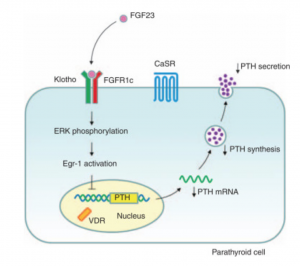
Fibroblast Growth Factor 23 (FGF-23) is a hormone primarily produced by osteocytes and osteoblasts in bones, playing a key role in regulating phosphate and vitamin D metabolism. It lowers serum phosphate levels by reducing phosphate reabsorption in the kidneys and suppresses the activation of vitamin D, thereby decreasing intestinal phosphate and calcium absorption. FGF-23 requires the co-receptor Klotho to bind to fibroblast growth factor receptors (FGFRs) and exert its effects, primarily in the kidneys. Dysregulation of FGF-23 is associated with several pathological conditions, making it a valuable biomarker for assessing phosphate balance and mineral metabolism.


image credit: https://www.kidney-international.org/


Why Measure FGF-23?

Measuring FGF-23 is essential for advancing research on phosphate metabolism, bone biology, and endocrine regulation. It provides insights into the mechanisms underlying phosphate homeostasis and the complex interplay between FGF-23, Klotho, and fibroblast growth factor receptors. FGF-23 is also a valuable marker for studying how mineral metabolism affects systemic processes, including cardiovascular function and aging. In experimental models, FGF-23 measurements help elucidate the effects of genetic mutations, dietary phosphate intake, and novel therapeutic interventions on phosphate regulation. Additionally, FGF-23 serves as a biomarker to explore the broader effects of mineral dysregulation on inflammation, oxidative stress, and metabolic disorders, enhancing our understanding of its role in health and disease.


Related Products:


*Use Promo Code: FGF25 to save 25% on your order!

We’re thrilled to announce that the MedFrontier Intact FGF23 Assay was featured in a recent publication! Details are available via the abstract and full-text access provided below.


Abstract

Multiple myeloma commonly manifests with symptoms arising from the involvement of various organs, particularly the bone and kidneys. In this report, we detail the case of a 44-year-old man who was diagnosed with multiple myeloma associated with reduced bone density. He exhibited clinical findings of osteomalacia due to Fanconi syndrome (characterized clinically by bone pain and proximal weakness and biochemically by elevated serum alkaline phosphatase, hypophosphatemia, hypouricemia, and glucosuria). With phosphate replacement, there was a notable improvement in bone pain, osteomalacia, and bone mineral density. Nevertheless, the patient continued to experience renal wasting of phosphate, uric acid, and glucose despite achieving remission from multiple myeloma for nearly 2 years. Our case highlights several important clinical features of myeloma-associated Fanconi syndrome, including the need to recognize this complication to appropriately treat the underlying bone disease while avoiding osteoclast inhibitors and the long-term persistence of the proximal renal tubulopathy despite achieving remission from myeloma and correction of osteomalacia.

Alireza Zomorodian, Naim M Maalouf, Long-term Evolution of Hypophosphatemia and Osteomalacia in a Patient With Multiple Myeloma, JCEM Case Reports, Volume 2, Issue 7, July 2024, luae137, https://doi.org/10.1210/jcemcr/luae137


If you have any questions about this product or any of our other offerings, contact us here.

Eagle Biosciences, Inc. is thrilled to announce a distribution partnership with Matriks Biotek, a leader in the development of ELISA kits for therapeutic drug monitoring!


About Matriks Biotek

Matriks Biotek was founded in 2002 by Prof. Haluk Ataoglu MD, PhD (Microbiology & Immunology). Their core business values include science, innovation, reliability, perfection and customer satisfaction. They were the first R&D company to globally commercialize ELISA kits for monitoring biological drugs, providing reliable tools for numerous research applications. With an offering of over 90 validated ELISA kits covering 36 biological drugs, Matriks Biotek’s product line includes quantitative free drug detection kits, qualitative anti-drug antibody detection kits, total antibody detection kits!

Key performance features of Matriks Biotek ELISA Kits:

  • Reliable Results: High recovery rates (85-115%) for robust results
  • Convenience: Minimal sample volume requirements (10-25 µl), even for small samples like mouse serum
  • Speed: Rapid processing times (70-140 minutes) for efficient workflows
  • Versatility: Broad sample compatibility (human, mouse, rat, and monkey serum or plasma)
  • Dynamic Range: Optimized for Cmax – Cmin (trough) values

Check out the full catalog, or reach out to us with any questions — we’d love to hear from you!

A recent publication employed the Bovine IgM ELISA Assay Kit to help measure the effects of commercial-scale heat treatments on milk proteins! This research provides key data on protein retention and functionality after processing. Check out the details and access the full findings below.


Abstract

Two pasteurization steps are often used in the preparation of whey protein concentrate (WPC) before evaporation into a dry product. The Pasteurized Milk Ordinance (PMO) in the United States requires that raw bovine milk be pasteurized using a process that meets minimum heat treatment requirements to achieve reductions in pertinent microorganisms. In addition, WPC produced from USDA-approved plants must comply with CFR Subpart B §58.809, which dictates that all fluid whey used in the manufacture of dry whey products shall be pasteurized before being condensed. These heat treatments are effective at inactivating the most thermally resistant bacterium, such as Coxiella burnetii; however, they can also alter milk proteins—inducing denaturation, aggregation and reduced bioactivity. Though the impact of thermal treatments on whey proteins has been examined, the specific influence of 2 high-temperature-short-time (HTST) pasteurization steps on the retention of proteins in WPC remains unknown. This study aimed to investigate the effect of commercial-scale HTST pasteurization of both raw milk and the resulting sweet whey on the products’ overall protein profile. Three distinct batches of raw milk (RM) and corresponding pasteurized milk (PM), the resulting whey (RW) and pasteurized whey (PW) produced at commercial scale were analyzed. Assessments of denaturation were conducted through solubility testing at pH 4.6 and hydrophobicity evaluation via anilinonaphthalene-1-sulfonic acid assay (ANS). Additionally, enzyme-linked immunosorbent assay (ELISA), PAGE (PAGE) and liquid chromatography tandem mass spectroscopy (LC-MS/MS) were employed to compare the retention of key bioactive proteins before and after each HTST pasteurization step. The percentage of soluble whey protein decreased from RM to PM and from RW to PW, but no significant differences were observed via hydrophobicity assay. ELISA revealed a significant reduction in key bioactive proteins, such as lactoferrin, immunoglobulin A and immunoglobulin M, but not immunoglobulin G, after HTST pasteurization of RM and RW. PAGE and LC-MS/MS revealed a significant decrease in the retention of lactoferrin and key milk fat globular membrane proteins, such as xanthine dehydrogenase oxidase/xanthine oxidase, lactadherin and fatty acid binding protein. Additionally, xanthine oxidase activity was significantly reduced after HTST pasteurization of milk and whey. This research helps to identify the limitations of the current processing techniques used in the dairy industry and could lead to innovation in improving the retention of bioactive proteins.

Haas, Joanna et al. Effects of high temperature short time (HTST) pasteurization on milk and whey during commercial whey protein concentrate production. Journal of Dairy Science, Volume 0, Issue 0. DOI: 10.3168/jds.2024-25493


If you have any questions about this kit or any of our other offerings, contact us here.