Human CDNF Mouse Monoclonal Antibody Clone 6G5

$610.00

The Human CDNF Mouse Monoclonal Antibody Clone 6G5 is intended for research use only and not for use in diagnostic or clinical procedures. Recombinant human CDNF protein produced using CHO-based Icosagen Cell factory Ltd. proprietary suspension cell line. Purified from cell culture supernatant.

SKU: 302-100 Categories: ,

Human CDNF Mouse Monoclonal Antibody Clone 6G5

The Human CDNF Mouse Monoclonal Antibody Clone 6G5 is For Research Use Only

Immunogen: Human CDNF
Uniprot ID: Q49AH0
Alternative Names: ARMETL1
Clone: 6G5
Clonality: Mouse monoclonal
Class: IgG1
Reactivity: Human, no reactivity with mouse CDNF
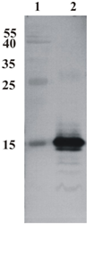
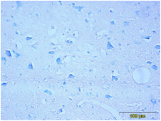
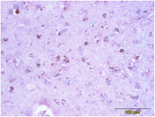
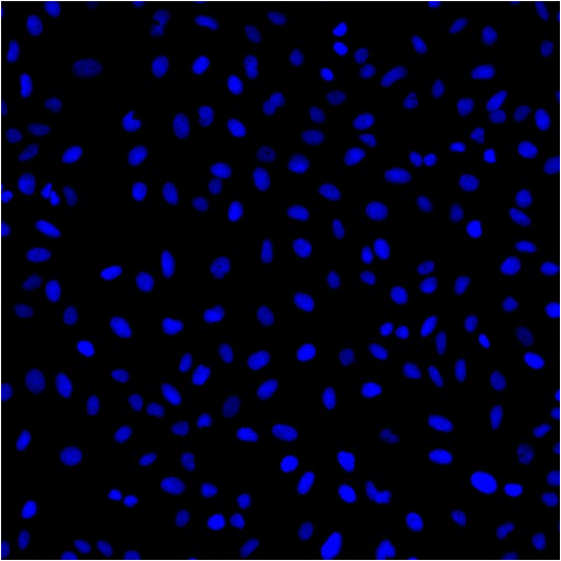
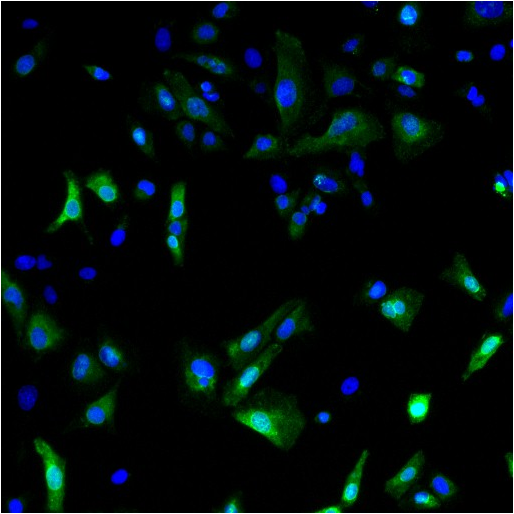
Application: ELISA, WB, IF, IHC
Purification: ELISA, WB, IF, IHC
Buffer: PBS pH 7.4, with 0.1% sodium azide
Concentration: 1 mg/ml
Unit Size: 100 µg
Related Products: Mono- and polyclonal antibodies to human CDNF.
Shipping: This product is shipped in non-frozen liquid form in ambient conditions
Storage: Store at –20 or -70 °C upon receipt. Divide antibody into aliquots prior usage. Avoid multiple freeze-thaw cycles as product degradation may result.
Alternative Names: Cerebral Dopamine Neurotrophic Factor


Protocol: ELISA – 1:5000 to 1:10 000;  WB – 1:1000 to 1:4000; IF – 1:100 to 1:300; IHC (on formalin-fixed, paraffin-embedded tissues, antigen retrieval) – 1:100 to 1:200; Monoclonal antibody working titer has to be established practically for each particular antigen and assay format

Background: CDNF is a trophic factor for midbrain dopamine neurons in vivo. It prevents the 6-OHDA-  (Lindholm et al. 20007; Voutilainen et al., 2011) and MPTP-induced degeneration (Airavaara et al., 2012) of dopamine neurons in rodent models of Parkinson’s disease. When administered after 6-OHDA or MPTP –lesioning it restores the dopaminergic function and prevents degeneration of dopamine neurons in substantia nigra pars compacta


Related Products

Human CDNF Mouse Monoclonal Antibody
Human CDNF Rabbit IgG Polyclonal Antibody
Human Recombinant CDNF Protein

Product Developed and Manufactured by Icosagen

Additional Information

Figure 1:
Human CDNF Mouse Monoclonal Antibody Clone 6G5


Western Blot testing of anti-CDNF monoclonal antibody (6G5). Line 1. PageRuler Prestained Protein Ladder (#SM0671 Fermentas); Line 2. Recombinant CDNF expressed into the supernatant of CHO cell culture medium.AB

Figure 2:

Human CDNF Mouse Monoclonal Antibody Clone 6G5

Human CDNF Mouse Monoclonal Antibody Clone 6G5


Immunohistochemistry testing of anti-CDNF monoclonal antibody 6G5. Analysis was performed using formalin-fixed paraffin-embedded human cerebral cortex tissue sections from Alzheimer’s disease patients. Tissue sections were boiled with sodium citrate buffer (pH 6) for antigen retrieval. Incubation with primary antibody at 5 µg/ml was performed overnight at 4°C. DAKO EnVisionTM Detection System, Peroxidase/DAB was used for visualization. Sections were counterstained with toluidine blue and mounted with Eukitt mounting medium. A. CDNF staining by monoclonal antibody 6G5; B. Negative staining without primary antibodyAB

Figure 3:
Human CDNF Mouse Monoclonal Antibody Clone 6G5

Human CDNF Mouse Monoclonal Antibody Clone 6G5


Immunofluorescence  detection of human CDNF expressed in U2OS cells. CDNF was visualized using anti-CDNF antibody clone 6G5 at 1 µg/ml. Goat ant-mouse AlexaFluor488 was used as secondary antibody. For nuclear staining DAPI was used. ArrayScan VTI platform (Thermo Scientific) was used for image acquisition (10x objective). Composite picture was generated using pseudocolors green for CDNF specific signal and blue for nuclei. A. CDNF-expressing U2OS cells; B. Negative control (non-transfected U2OS cells)

Package Inserts


Please note: All documents above are for reference use only and should not be used in place of the documents included with this physical product. If digital copies are needed, please contact us.