EZ4U ELISA Assay Kit

$295.00

The Eagle Biosciences EZ4U Cell Proliferation Assay is for research use only and not to be used in diagnostic procedures.

SKU: BI-5000 Category:

EZ4U ELISA Assay Kit

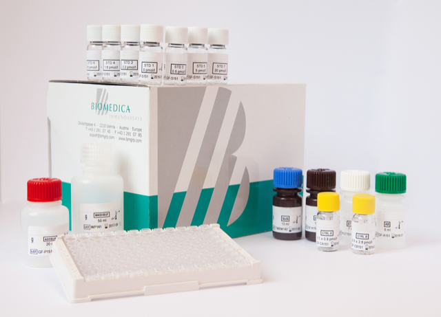
EZ4U ELISA Assay Kit Developed and Manufactured in Austria by Biomedica

Method: Cell proliferation and cytotoxicity assay, 10 x 96 tests, method based on the reduction of tetrazolium salt to colored formazan
Incubation Time: 2 – 5 hours, depending on the metabolic capacity of the cells
Sample Type: Cell culture supernatant
Sample Size: 200 µl / test
Alternative Names: Cell proliferation and cytoxicity assay
For Research Use Only


Assay Overview

The EZ4U assay is a two to five-hour test for the measurement of cell proliferation and cell toxicity. The assay employs non-toxic tetrazolium salts, which are reduced to colored formazan. As this reduction process requires functional mitochondria, which are inactivated within a few minutes after cell death, this method provides an excellent tool for the discrimination between living and dead cells.


Assay Principle

The EZ4U ELISA Assay Kit set-up is performed in the same manner as the standard 3H-thymidine incorporation method. Instead of pulsing with tritiated nucleotide, 20 µl of dye solution is added to 200 µl sample. The incubation time is dependent on the metabolic capacity of the cells. Usually two to five hours of incubation at 37°C are sufficient to yield a significant increase in color intensity. As different cells vary in their ability to convert the yellow colored tetrazolium compound to its red formazan derivative, we recommend testing every new cell-line’s metabolic capacity as described in the figure below.
After incubation, the plate is removed from the incubator and gently mixed by tipping the plate towards all four sides. To avoid increased standard deviations, the plate must be shaken before reading the optical density.
The absorbance is measured on a microplate-reader set at 450 nm or 492 nm with 620 nm as a reference wavelength. The reference absorbance at 620 nm (or any wavelength between 620-690 nm) is used to correct for nonspecific background values caused by cell debris, fingerprints, or other potential interferences. However, the reference may be omitted without significant changes in the accuracy of the assay.

Related Products

Total Soluble Neuropilin-1 ELISA
FGF23 C-Terminal ELISA Kit
Human MCP-1 ELISA Assay Kit

Additional Information

Assay Background


Proliferation assays are widely used in cell biology for the study of growth factors, cytokines, nutrients and for the screening of cytotoxic or chemotherapeutic agents. There are several ways to determine the number of cells either by microscopic inspection, or by the use of an electronic particle counter, indirectly by measuring the incorporation of radioactive precursors, quantitating total protein with chromogenic dyes, or by measuring metabolic activity of cellular enzymes. The most common assay for cell proliferation is the incorporation of 3H‑thymidine into cellular DNA. The 3H-thymidine assay is, however, labour intensive as it requires the removal of excess, unincorporated label by using some method of cell harvesting before measurement. In 1956, the first paper was published on the use of tetrazolium salts as indicators of cell viability. The method was based on the finding that living cells are capable to reduce slightly or uncoloured tetrazolium salts into intensely coloured formazan derivatives. This reduction process requires functional mitochondria, which are inactivated within a few minutes after cell death. This method therefore provides an excellent tool for the discrimination of living and death cells. However, the early tetrazolium salts did have some disadvantages, such as the insolubility of the resulting formazan products. Time and labour consuming resolubilisation procedures were necessary, including repipetting and mixing, or the application of hazardous solubilisers. This necessary post assay treatment, however, irreversibly terminated cell proliferation and thus made it impossible to prolong incubation in order to achieve an increase in sensitivity or continue cell culture. These inconveniences led to the development of non-toxic tetrazolium salts which yield soluble reduction products. Although the assay procedure was made easier by these soluble dyes, in practice the use was limited due to the instability of the formazan dye and a relatively low absorbance of the end product as compared to the classical MTT assay.
The BIOMEDICA research department has solved both problems and created an easy to use, rapid and reliable non-isotopic cell proliferation assay. For convenience, we have made it highly compatible with the standard thymidine incorporation assay. Therefore, no changes are required in the setup of the test and in the “labelling” procedure. Furthermore, there is no need for the removal of culture medium before or after the addition of the chromogenic substrate and neither solubilisation nor harvesting procedures are necessary. The work performed by BIOMEDICA resulted in an assay which combines the best of the thymidine and MTT methods, namely: accuracy, speed, reliability and ease of use. Also, according to our data achieved so far, the chromophore appears to be non-toxic. A double labelling with EZ4U and a radioactive nucleotide to obtain more information about cell viability and DNA content is now feasible.

Assay Principle


  • The assay set-up is performed in a manner similar to the standard 3H-thymidine incorporation method.
  • Instead of pulsing with tritiated nucleotide, 20 µl of dye solution is added to 200 µl sample.
  • Incubation time is dependent on the metabolic capacity of the cells.
  • Usually 2 to 5 hours of incubation at 37°C are sufficient to yield a significant increase in color intensity.
  • As different cells vary in their ability to convert the yellow colored tetrazolium compound to its red formazan derivative, we recommend testing every new cell-line’s metabolic capacity as described in Fig.2.
  • After incubation, the plate is removed from the incubator and gently mixed by tipping the plate at all four sides.
  • To avoid increased standard deviations, the plate must be shaken before reading the optical density.
  • The absorbance is measured by a microplate-reader, set at 450 nm or 492 nm with 620 nm as a reference. The reference absorbance at 620 nm (or any wavelength between 620-690 nm) is used to correct for nonspecific background values, caused by cell debris, fingerprints, or other potential interferences. However, the reference may be omitted without significant changes in the accuracy of the assay.

Package Inserts


Please note: All documents above are for reference use only and should not be used in place of the documents included with this physical product. If digital copies are needed, please contact us.

Product Citations