What is it AgarSqueezer intended for?

AgarSqueezer is a microscope slide chamber equipped with a molded agar-based compression system. Use it to assess cell response to short and long-term mechanical confinement within a physiological rigidity range.

It is very helpful if you want to analyze how your cells will react if you squeeze them for a prolonged period. Or if you want to study how mechanical confinement affects drug cell resistance. And if you want to perform immunostaining in situ.


Kit Description:

You can choose among 2 kits:

  • The “1 AgarSqueezer kit” contains 1 AgarSqueezer device
  • The “2 AgarSqueezers kit” contains 2 AgarSqueezers devices.

In addition to the device, each kit contains also:

  • 1 x insert to hold up to 2 AgarSqueezers on the microscope stage
  • 1 x 16G flat cut needle to make holes in the agar gel, facilitating diffusion of culture medium or drugs during the experiment
  • 1 x 20G flat cut needle (same)

For users who also need a wafer to mold agarose, contact us to learn about the four different heights we offer.


Features

  1. Tunable stiffness in a physiological range [1-150] kPa – Use of agarose as a cheap and biocompatible polymer.
  2. Open access to the reservoir – Possibility to add drugs, and reagents. Easy medium renewal.
  3. Autoclavable and reusable systems.
  4. Compatibility with multiple microscopy techniques – Confocal, spinning, super-resolution. Open access for microscope objectives. Use of optical glass coverslip to make cells grow.
  5. Easy to recover coverslip with cells for subsequent molecular analysis – FACS, qPCR, Western-Blot, Immunoflourescence (possible in situ).
  6. Long-term analysis of the cell adaptation to confinement – Up to several days, for time-lapse studies.
  7. Study of the specific impact of mechanical loads on the biology of cells – Gas permeability of the system allows to get rid of the hypoxia conditions.
  8. Easy to assemble and disassemble the system

For more information about this product or any others from the Microscopy line, contact us here.

Eagle Biosciences is excited to announce the newest product series for Host Cell Protein Detection!

Host cell proteins (HCPs) are a major class of impurities produced during biotherapeutic manufacturing. They must be removed from the final drug product to both assure patient safety and maintain drug efficacy. Our wide range of Host Cell Protein Detection Kits are easy to use and highly sensitive.


What are HCPs and why must they be removed from biologic drugs?

HCPs are proteins produced or encoded by the host organisms used to produce recombinant therapeutic proteins. Genetic engineering allows the host organism cells to be transformed to produce a protein of interest. During the recombinant protein production, host cells also coproduce proteins related to the normal cell functions such structural proteins, as well as proteins required for normal cellular growth and function, and vary in both number and concentration depending on the chosen host species and the manufacturing process being used. In general, apart from the therapeutic protein of interest, all endogenous proteins co-expressed by the host cells are called host-cell proteins.

Why must HCPs be removed from biologic drugs?

HCPs must be removed from the final biotherapeutic product to avoid adverse effects. Almost all HCPs carry safety risks as foreign proteins due to the potential to elicit immune response in humans (e.g, cytokine storm). In addition, some HCPs can also act to enhance the immune response to a drug product. Certain HCPs can also affect drug product stability and efficacy if not adequately removed or inactivated.

How are HCPs detected?

ELISAs are widely used for detecting HCPs, where they are generally configured in a sandwich assay format for improved specificity. In this scenario, a microplate-bound antibody is used for analyte capture, then a second analyte-specific antibody (that binds a different epitope on the target molecule) is added to enable detection. By incorporating a reference standard (e.g., a purified protein) into the assay design, it is possible to quantify the analyte of interest and confirm that its concentration meets regulatory requirements. Advantages of ELISA are that it is sensitive and compatible with high sample throughput – key considerations for biopharmaceutical manufacturing.


If you have any questions about these products or any of our other offerings, contact us here.

What is it SpheroRuler intended for?

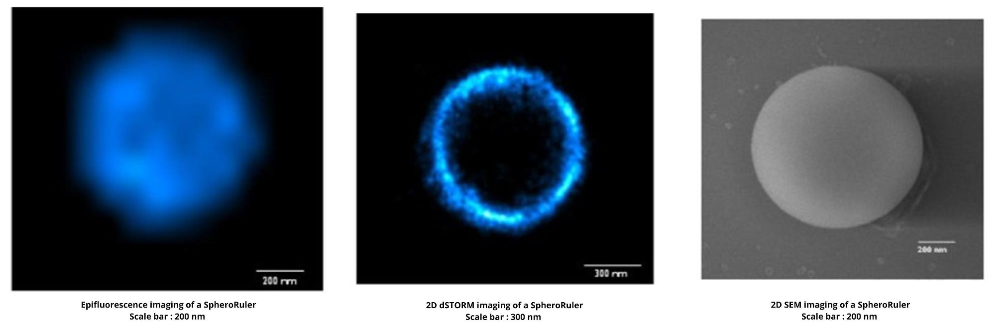
SpheroRuler is a monodispersed suspension of 1µm diameter spheres coated with 647-fluorophores giving a stable blinking in SMLM microscopy. Their consistent size and geometry make them very practical and reliable standards to assess the accuracy of your x-y or z measurements, 3D reconstruction methods or your image quality. Monodispersed and immobile in solution, they can also be used as rulers, for drift correction or as guides to help localize features on your biological samples.

SpheroRuler is great for dSTORM calibration experiments because you can use it on your preferred support and buffer. And because you get confident with the new biological structure to image. SpheroRuler also produces simple shapes that are easy to study when you are a dSTORM beginner!


Features

  1. Stable Blinking – Suited for use in SMLM microscopy
  2. Consistent Size – Thoroughly characterized by electron microscopy
  3. Spherical geometry – with sharp and thin edges
  4. Immobile – Suitable for drift correction applications
  5. Bio-compatible – Resuspended in an aqueous buffer and usable along biological samples such as cells or tissue sections
  6. Easy-to-Spot – A high fluorescence intensity for practical use as demo or training tools


For more information about this product or any others from the Microscopy line, contact us here.

What is Stencell intended for?

Stencell is used notably for wound-healing assay & immunocytochemistry experiments. It is very helpful when you are working with super expensive reagents or very rare cell lines, and when you want to test various experimental hypothesis.

Stencell for wound-healing experiments


Stencell Designs

Each order is composed of 2 sheets of 50 identical stencils (=100 stencils). Upon receipt of your purchase order, you will be able to choose either the same design for your 2 sheets, or 2 different designs for your 2 sheets (1 per sheet of 50).

    • Solo: 1 circular well – Diam. 12 mm
    • Quartet: 4 circular wells – Diam. 3 mm
    • Nonet: 9 circular wells – Diam. 3 mm
    • Presto: 2 oblong wells spaced by 0.35 mm
    • Allegro: 2 oblong wells spaced by 0.62 mm


    Features

    1. Design your PDMS – Our design process offers total freedom to turn your idea into a PDMS.
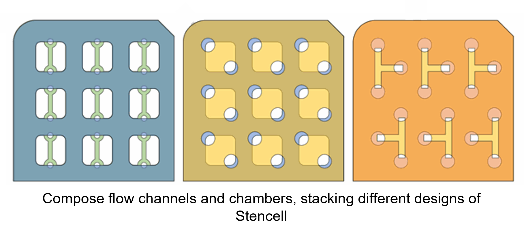
    2. Stack and Play – Compose flow channels and chambers, stacking different designs of Stencell.
    3. Stick it to a variety of culture labware – So far, it has been successfully stuck onto the following: glass slides, plastic dishes, Transwell(R) inserts, Polyacrylamide gels.
    4. Remove it – Stencell is not glued. It can then be removed to trigger cell migration, switch from flow chamber to open window. (Very useful for wound healing experiments)
    5. It is compatible with imaging – These thin sheets of silicone are fully transparent, without autofluorescence.
    6. Parallelized and standardized experiments – One multiwell Stencell can perform several experimental conditions.
    7. It saves samples and reagents – Only a few microliters are required.


    Key Conditions of Success

    • When you remove the inner elements of your Stencell, use 2 tweezers: 1 to remove and the other 1 to press close to the junction in order to avoid breaking the Stencell. Alternatively, you can cut the junctions with a scalpel.
    • When you put the Stencell on top of your culture substrate, softly remove the bubbles and stick the Stencell, patting it with tweezers.
    • Depending on the substrate hydrophobicity, the droplet might not completely fill the well. You can:
      • Either fill the well with the standard volume. Then, help the droplets stick to the Stencell by connecting the liquid next to the Stencell walls using the pipette tip.
      • Or, you may add an excess volume of liquid (e.g. 30 μL) and remove it afterwards (e.g. 10 μL).
    • But: do not overfill (e.g. > 30 μL) the wells, otherwise two droplets may merge.
    • When you put your substrate and cells in the incubator. Wait for 1 to 2 hours until cells spread and fully occupy the windows space. If you need to wait longer, be sure that the droplets do not completely evaporate.

    For more information about this product or any others from the Microscopy line, contact us here.

What is it Stampwell intended for?

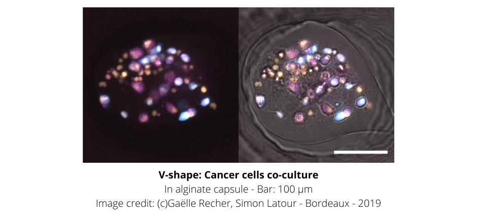
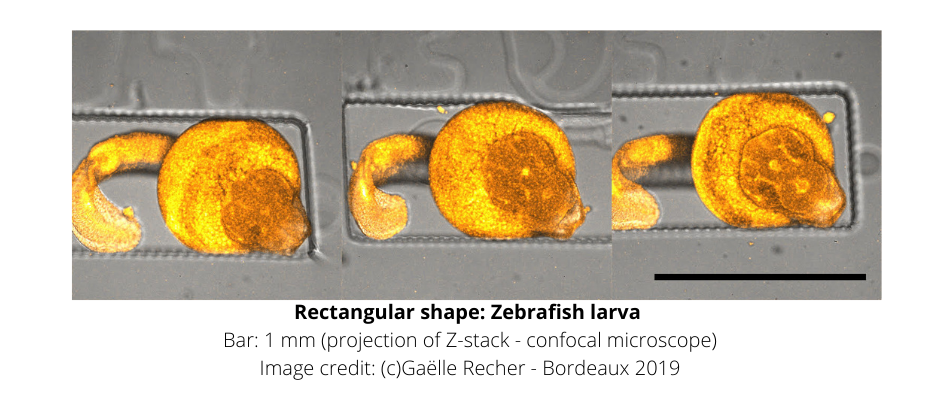
Stampwell is highly helpful if you have tons of 3D samples to image. The V-shape Stampwell is used to image them (2 different sizes). And the Rectangular Stampwell is designed to image Zebrafish or Medaka embryos. It avoids spending hours to locate your sample in the dish. It prevents them from slipping from one well to another when you move your dish from the incubator to the microscope. And it allows to image and observe how your biological samples grow over several weeks.


3 Shapes of wells to choose freely and best fit your experiments:

  • V Shape: 7*6 pins in V-shape – Diameter 26mm – Exists in 2 different sizes: 300 or 500u
  • Rectangular:7*5 rectangular – Diameter 26mm
  • “My Shape”: design your wells, adapted to your samples and your experimental needs!


Features of Stampwell

  1. Cross-platform – Compatible with most 35 mm dishes and 6-well plates.
  2. Reusable – Use your stamp dozens of times.
  3. Medium Culture Conditioning – U Shape: the design of a groove increases the culture medium conditioning and cell aggregates survival.
  4. Long-term imaging over weeks with V-shape and Rectangular – the sample just lays on the bottom of the well and can freely grow within it.
  5. Safe transport of 3D objects – V-shape and Rectangular keep your samples at the bottom of the wells, even when the dish is upside down.
  6. Automatized multi-position image acquisition V-shape and Rectangular – provide a constraint-free but stable and parallelized immobilization of the samples.
  7. Customize your shape – My-Shape is your own design and number of wells. Contact us for details.


For more information about this product or any others from the Microscopy line, contact us here.

What is Everspark intended for?

Everspark is a hassle-free buffer used to decouple the preparation of your samples and their imaging. Use it if you need to prepare your samples in your lab and image them in your core facility 3 to 4 weeks later. Everspark is also compatible with imaging that uses red, far-red and green dyes (eg. AF488). Different colors can be used either at the same time or one after the other.


Features

  1. High stability over time for repeated measurements – One mounted sample is stable over 3 to 4 weeks and can be imaged multiple times
  2. Multicolor (including green) – Compatible with green, red, and far red dyes (JF646, JF549, AF647, DL550, CF568, DL650, CF680, SulfoCy5 and mEos2)
  3. Ready to Use – Can be used directly from the vial
  4. Up to 6 months performance – Individualized packaging for optimal longevity
  5. Techniques – PALM, dSTORM, HILO, TIRF pattern
  6. Customizable – Choose the MEA concentration, pH, optical index, or even additives


Use Method:

  • Rinse the coverslip with sterile water
  • Discard water in excess
  • Prepare sealant: 1 volume of sealant catalyst + 1 volume of sealant basis in a micro-dish – do not mix
  • Open the Everspark tube and pipette right away the desired volume onto a Wilco glass bottom dish
  • Flip the coverslips face towards the slide on the cavity filled with Everspark without making bubbles
  • Absorb excess of Everspark buffer on the sides with a kimwipe
  • Mix sealant
  • Seal quickly with the reconstituted sealant
  • Wait for the sealant to polymerize on the slide before moving the slide
  • Discard the open tube of Everspark buffer (cannot be used for another slide, once already opened)
  • Gently clean the coverslip with 70% ethanol
  • Perform regular dSTORM acquisition (best to wait a couple of hours)
  • Repeat dSTORM acquisition 24 hours up to 3 weeks on the same slide providing no air bubbles were present
  • Store the slides at 4°C and in the dark before repeating dSTORM acquisition on the same slide (up to 3 weeks)

For more information about this product or any others from the Microscopy line, contact us here.

What is Chitozen intended for?

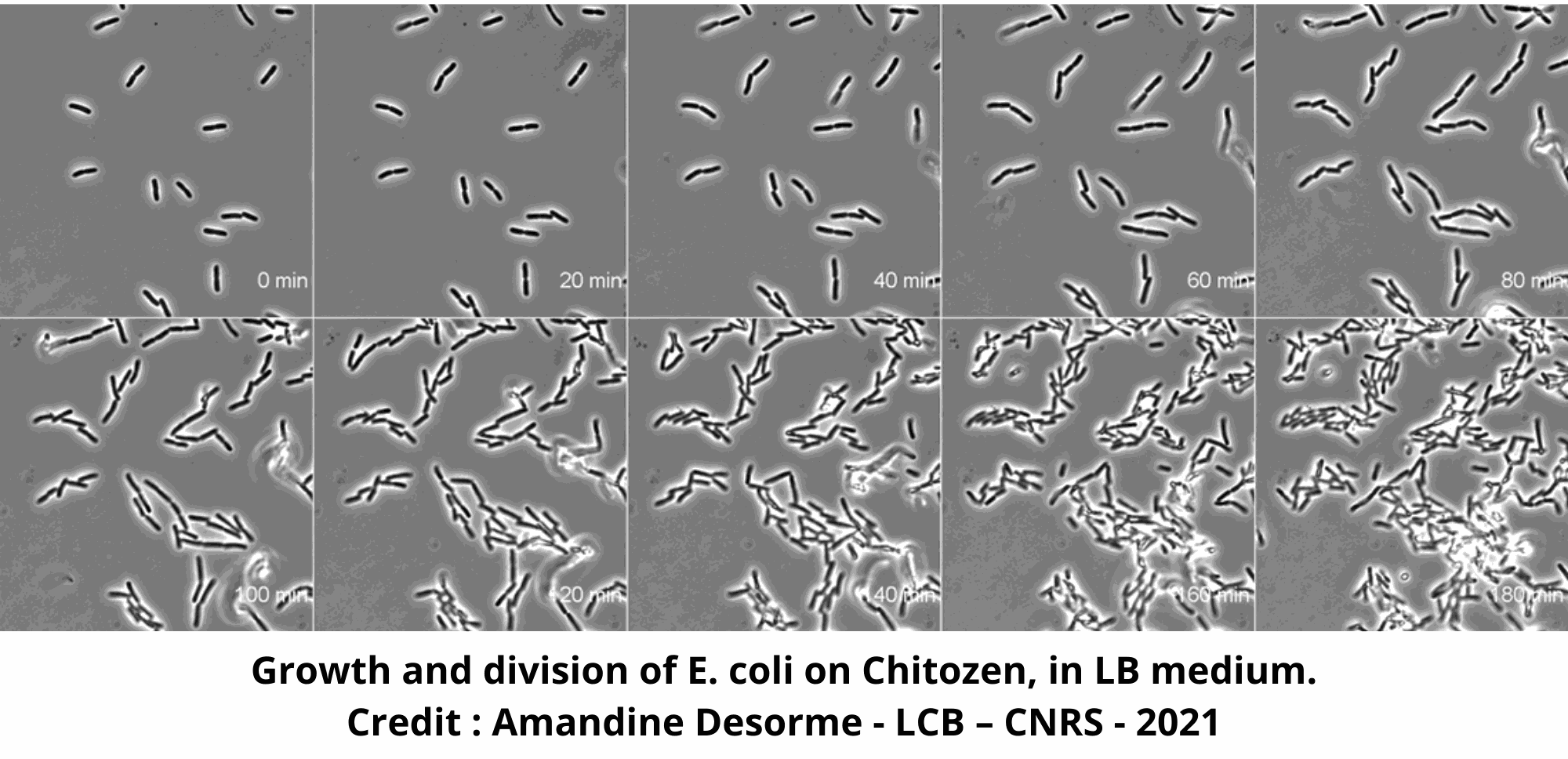
Chitozen is used to image bacteria both still and alive under the microscope, or it can be used to perform long-term imaging of bacteria. It can also be used if you want to change the growth condition (e.g. antibiotics, chemicals, inhibitors) during the experiment and directly observe, in real-time, the bacteria new comportment under the microscope.

E.coli monolayers on Chitosan


For what bacteria is Chitozen used for?

Chitozen has been tested with the following bacteria: E. coli, Vibrio cholerae, Myxococcus xanthus, Mycobacterium smegmatis, Bacillus subtilis, Pseudomonas aeruginosa and Pseudomonas fluorescens. More bacteria being currently tested.

Growth of E.coli on Chitozen


Simplified Protocol


Features

  1. Full compatibility with most of your conditions of experiments – Size: the coverslip dimension (25x75mm) is compatible with the most common available sticky slides and microscope stages; it is also compatible with advanced microfluidic techniques, nanolithography
  2. 6 independent channels – Either perform up to 6 experiments at the same time or use 1 channel one day, and the others later
  3. Ready and fast – Assemble it within 2 minutes, use it the same day as its prepared
  4. Long lasting – A bench-stable surface coated with chitosan, the most efficient way to immobilize your bacteria on a microscope coverslip; storable for 2 months once assembled


For more information about this product or any others from the Microscopy line, contact us here.

What is Actiflash intended for?

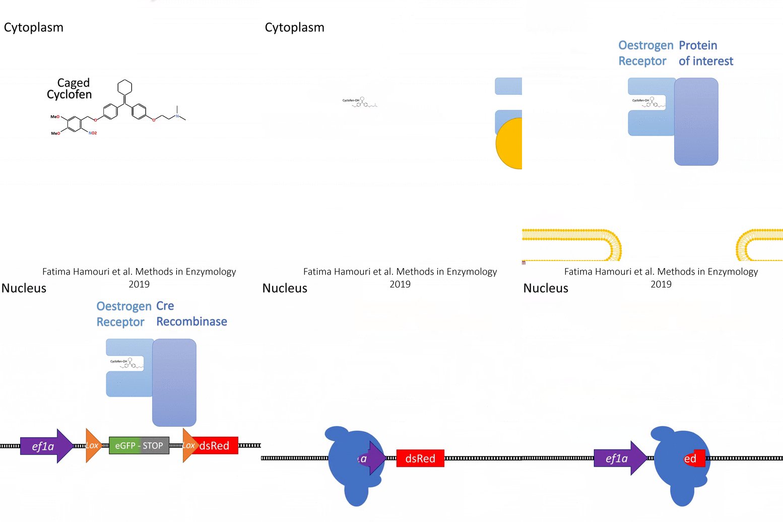
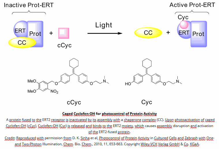
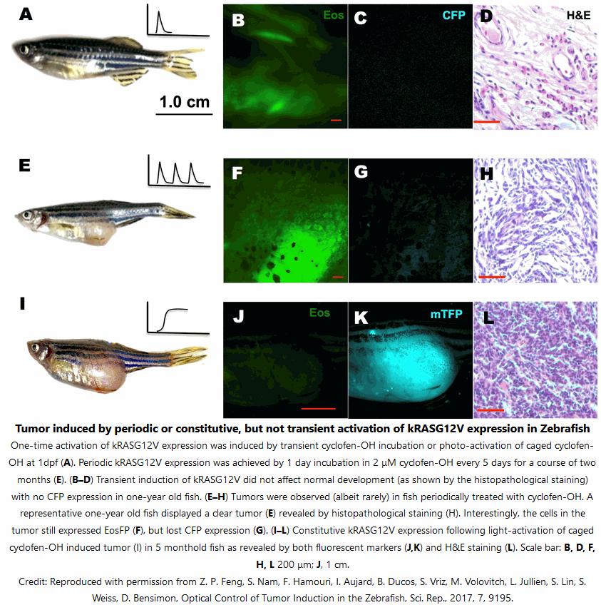
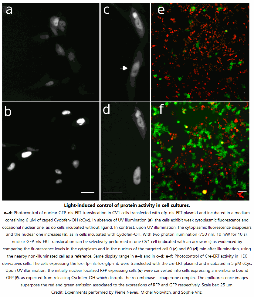
Actiflash is used to convert your inducible ERT model into a photo-inducible one. Use it if you want to control transcription (using Gal4-UAS) or induce recombination (using Cre-lox) in space and/or time for in-vivo cell tracking experiments and more. Actiflash is for research use only


Features

    1. Wide applicative scope – Technology capitalizing on the versatile use of Tamoxifen-OH for controlling functions of multiple types of proteins.
    2. Simple conditioning – Caged Cyclofen-OH is cell-permeant and can be added either in the external medium or directly injected for conditioning.
    3. Excellent chemical stability – Caged Cyclofen-OH does not generate any basal activation of protein function and it benefits from an excellent temporal resolution upon uncaging.
    4. Favorable wavelength ranges for uncaging – Uncaging requires either UV-A light or a strong IR laser. Visible light is inactive, which facilitates the experiments with biological samples.
    5. Photochemical stability – Caged Cyclofen-OH liberates Cyclofen-OH, which is photostable in contrast to Tamoxifen-OH encountering photodegradation under illumination.


Method of Use:

Calibration of the Actiflash concentration
It is advised to first establish the extent of phenotype sought for as a function of the Tamoxifen-OH concentration. Then the concentration of Actiflash used for sample conditioning is fixed at Tamoxifen-OH concentration causing 100% of the desired phenotype (in general 3-5 μM in cultured cells and zebrafish embryos).

Conditioning protocol
Incubate your samples in a serum-free medium for 90 mins, away from light.

Photoactivation
Illumination of Actiflash may be performed with UV (325-425nm range) light or multiphoton excitation (at 750 and 1064 nm with two- and three-photon excitation, respectively) to release Cyclofen-OH. You can use either benchtop UV lamps or light sources installed on microscopes.

The calibration of the photoactivation
The objective is to provide enough photons to exhaust the conversion of the Actiflash but without generating detrimental side-effects on the biological sample. Simply analyze the phenotype recovery with decreasing illumination duration. Then determine the shortest illumination duration leading to 100% uncaging of Actiflash.


5 Key Conditions of Success

  1. Actiflash is provided as a powder. Resuspend it with DMSO (warning: no water).
  2. Only unfreeze the aliquots you need for your experiment.
  3. Carefully calibrate the photoactivation of Actiflash (concentration for conditioning, power of the light source, geometry and duration of illumination). Then always work under the same conditions.
  4. Use a serum-free medium during the incubation step of the cell lines.
  5. In the dark, just before photoactivation, wash your samples (unnecessary for two-photon laser experiments).


For more information about this product or any others from the Microscopy line, contact us here.

At this time it is unknown for how long antibodies persist following infection or vaccination, and if the presence of antibodies confers protective immunity. Antibodies to SARS-CoV-2 are generally detectable in blood several days after initial infection or vaccination, although the duration of time antibodies are present post-infection or post-vaccination is not well characterized. Individuals may have detectable virus present for several weeks following seroconversion.

The Cov19 FluoBolt-DAT test, manufactured in Austria by Fianostics GmbH, is a duplex antibody test that is intended to identify individuals with an adaptive immune response to SARS-CoV-2, indicating recent or prior infection or vaccination. There is a high demand to know how protected populations are after COVID-19 onset or vaccination, this test is the next step in identifying immunity.


How the Cov19 FluoBolt-DAT Test Works

When performing the assay, 10 µl of a patient serum or plasma sample are simultaneously mixed with 50 µl of two different fluorescence-labeled tracer antibodies against the S1RBD and against the NC protein and incubated in our special fluorescence-enhancing microtiter plate for 60 minutes. By measuring at 2 different wavelengths, their displacement by homologous antibodies from the patient sample is determined simultaneously. The signals at one wavelength provide information about the concentration of anti-S1RBD antibodies. The signals at the other wavelength indicate the concentration of anti-nucleocapsid antibodies.

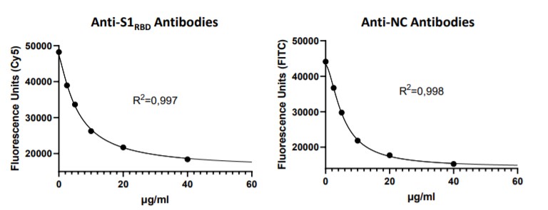
By using the supplied calibrators, a calibration curve for the anti-S1RBD and the antiNC antibodies is constructed, and the antibody concentration of a patient sample is read from it. With just a single measurement, this results in a quantitative determination of these
two antibody species against SARS-CoV-2, which represent the majority of the immune response and may protect against infection and illness.

Cov19 FluoBolt-DAT Test Standard Curves
For measurement, any commercially available fluorescence microtiter plate reader can be used.

Click here to learn more about the Cov19 FluoBolt-DAT test.


If you have any questions about this kit or our other offerings, contact us here.

IHC Antibodies

Eagle Biosciences, Inc. is excited to announce a new line of monoclonal IHC Antibodies. These anti-human antibodies are intended to be used to identify the presence of associated antigens in tissue samples. This identification should be made using light microscopy in formalin-fixed, paraffin-embedded (FFPE) tissue sections using immunohistochemistry test methods.  The use of these antibodies has been show to be an aid in the identification of neoplastic tissue, within the context of antibody panels. These IHC Antibodies are available in two formats. We offer a concentrated formula at volumes of 0.1 mL and 1.0 mL, as well as a prediluted formula at a volume of 7 mL.

Find a list of the popular and unique markers that we are now offering below.


IHC Antibodies

Monoclonal Mouse Anti-Human SOX-10 Antibody
Monoclonal Rabbit Anti-Human PD-L1 Antibody
Monoclonal Mouse Anti-Human HER2/neu Antibody
Monoclonal Rabbit Anti-Human Estrogen Receptor Antibody
Monoclonal Mouse Anti-Human BRAF V600E Antibody
Monoclonal Mouse Anti-Human p63 Antibody
Monoclonal Mouse Anti-Human p16 INK4A Antibody
Monoclonal Mouse Anti-Human MLH1 Antibody
Monoclonal Mouse Anti-Human MDM2 Antibody
Monoclonal Rabbit Anti-Human Ki-67 Antibody
Monoclonal Mouse Anti-Human IDH1 R132H Antibody


If you have any questions about these products or any of our other offerings, contact us here.