Mouse sRANK Ligand ELISA Assay

$2,065.00

The Mouse sRANK Ligand ELISA Assay Kit is intended or the quantification of Mouse soluble Receptor Activator of NFkB Ligand in serum, plasma, cell culture supernatants. The Eagle Biosciences sRANK Ligand ELISA Assay Kit is for research use only and should not be used for diagnostic procedures.

Mouse sRANK Ligand ELISA Assay

Mouse sRANK Ligand ELISA Assay is For Research Use Only

Size: 1 x 96 wells
Sensitivity: 15 pg/ml
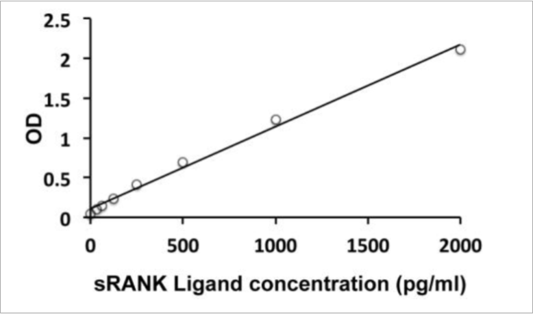
Dynamic Range: 31.25-2000 µg/ml
Incubation Time: 4 hours
Sample Type: Mouse Serum, Plasma, Cell Culture Supernatants
Sample Size: 100 µl
Alternative Names: Soluble RANK Ligand, sRANKL, TNFSF11, TRANCE


Assay Principle

The Mouse sRANK Ligand ELISA Assay employs the quantitative sandwich enzyme immunoassay technique. An antibody specific for sRANK Ligand / TNFSF11 / TRANCE has been pre-coated onto a microtiter plate. Standards or samples are pipetted into the wells and any sRANK Ligand / TNFSF11 / TRANCE present is bound by the immobilized antibody. After washing away any unbound substances, a biotin-conjugated antibody specific for sRANK Ligand / TNFSF11 / TRANCE is added to each well and incubate. Following a washing to remove unbound substances, streptavidin conjugated to Horseradish Peroxidase (HRP) is added to each microplate well and incubated. After washing away any unbound antibody-enzyme reagent, a substrate solution (TMB) is added to the wells and color develops in proportion to the amount of sRANK Ligand / TNFSF11 / TRANCE bound in the initial step. The color development is stopped by the addition of acid and the intensity of the color is measured at a wavelength of 450nm ±2nm. The concentration of sRANK Ligand / TNFSF11 / TRANCE in the sample is then determined by comparing the O.D of samples to the standard curve.


Related Products

iLite RANKL Assay Ready Cells
Free soluble RANKL ELISA
Mouse Soluble alpha-Klotho ELISA

Additional Information

Assay Background


This gene encodes a member of the tumor necrosis factor (TNF) cytokine family which is a ligand for osteoprotegerin and functions as a key factor for osteoclast differentiation and activation. This protein was shown to be a dentritic cell survival factor and is involved in the regulation of T cell-dependent immune response. T cell activation was reported to induce expression of this gene and lead to an increase of osteoclastogenesis and bone loss. This protein was shown to activate antiapoptotic kinase AKT/PKB through a signaling complex involving SRC kinase and tumor necrosis factor receptor-associated factor (TRAF) 6, which indicated this protein may have a role in the regulation of cell apoptosis. Targeted disruption of the related gene in mice led to severe osteopetrosis and a lack of osteoclasts. The deficient mice exhibited defects in early differentiation of T and B lymphocytes, and failed to form lobulo-alveolar mammary structures during pregnancy. Two alternatively spliced transcript variants have been found. Cytokine that binds to TNFRSF11B/OPG and to TNFRSF11A/RANK. Osteoclast differentiation and activation factor. Augments the ability of dendritic cells to stimulate naive T-cell proliferation. May be an important regulator of interactions between T-cells and dendritic cells and may play a role in the regulation of the T-cell-dependent immune response. May also play an important role in enhanced bone-resorption in humoral hypercalcemia of malignancy (By similarity). Induces osteoclastogenesis by activating multiple signaling pathways in osteoclast precursor cells, chief among which is induction of long lasting oscillations in the intracellular concentration of Ca (2+) resulting in the activation of NFATC1, which translocates to the nucleus and induces osteoclast-specific gene transcription to allow differentiation of osteoclasts. During osteoclast differentiation, in a TMEM64 and ATP2A2-dependent manner induces activation of CREB1 and mitochondrial ROS generation necessary for proper osteoclast generation.


Assay Procedure

  1. Remove excess microplate strips from the plate frame, return them to the foil pouch containing the desiccant pack, and reseal it.
  2. Add 100 μl of standards, samples and zero controls (standard diluent buffer) into wells. Incubate for 1.5 h at 37°C.
  3. Aspirate each well and wash, repeating the process four times for a total five washes. Wash by filling each well with 1× Wash Buffer (350 μl) using a squirt bottle, manifold dispenser, or autowasher. Complete removal of liquid at each is essential to good performance. After the last wash, remove any remaining Wash Buffer by aspirating, decanting or blotting against clean paper towels.
  4. Add 100 μl 1X Antibody conjugate into each well. Cover wells and incubate for 1 hour at 37°C.
  5. Aspirate each well and wash as step 3.
  6. Add 100 μl of 1X HRP-Streptavidin solution to each well. Cover wells and incubate for 30 minutes at 37°C
  7. Aspirate each well and wash as step 3.
  8. Add 100 μl of TMB Reagent to each well. Incubate for 15 minutes at 37°C in dark.
  9. Add 100 μl of Stop Solution to each well. The color of the solution should change from blue to yellow.
  10. Read the OD with a microplate reader at 450nm immediately.

Typical Standard Curve


Mouse sRANK Ligand ELISA

Package Inserts


Please note: All documents above are for reference use only and should not be used in place of the documents included with this physical product. If digital copies are needed, please contact us.

Product Citations