

How does it work?
GlowMito provides a non-toxic solution to quickly visualize the entire mitochondrial network in live samples.
GlowMito quickly penetrates cells and produces a bright & stable red labeling of mitochondria without inducing cell toxicity or altering mitochondrial functions. produces a bright, red labeling of the entire mitochondrial network, including those with reduced potential. It is perfectly suitable for:
- Live imaging: study of mitochondrial dynamics (velocity, localization), structural changes, multiplexing (potentiometric dyes, calcium signaling probes, etc)
- Downstream analysis: flow cytometry, oxygraphy, etc
We do not recommend its use to measure mitochondrial mass or volume density.
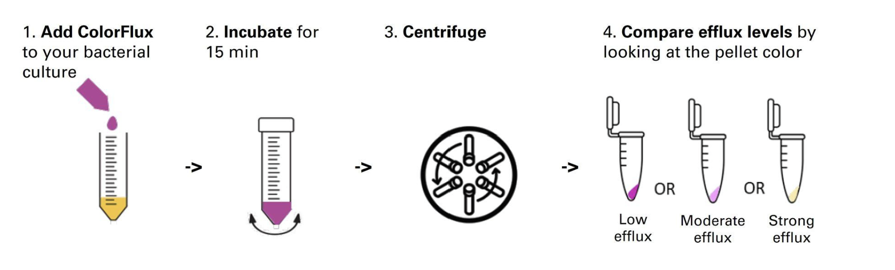
Simplified Protocol
Frequently Asked Questions
How was mitochondrial specificity of GlowMito verified?
The strong ability of GlowMito to specifically target mitochondria has been meticulously checked by co-localization studies. More generally, the ability of lipophilic cations to specifically target mitochondria is already well-established and conjugating molecules to lipophilic cations is a commonly used method to develop mitochondria-targeted compounds.
Which assays can GlowMito be used for?
GlowMito produces a bright, red labeling of the entire mitochondrial network, including those with reduced potential. It is perfectly suitable for:
- Live imaging: study of mitochondrial dynamics (velocity, localization), structural changes, multiplexing (potentiometric dyes, calcium signaling probes, etc)
- Downstream analysis: flow cytometry, oxygraphy, etc
We do not recommend its use to measure mitochondrial mass or volume density.
Which types of samples has GlowMito been used with?
So far, GlowMito has been successfully used to label mitochondria in the following biological samples:
- Human cells: HEK293, HeLa, MCF-7, MDA-MB-231, HMLE, UACC-62, U2-OS, Gli36, HAEC, SH-SY5Y, A-172, A549, patient-derived skeletal muscle cells & primary smooth muscle cells
- Monkey cells: COS-7
- Mice cells: primary cortical neurons
- Tissues: hiPSC-derived heart tissues & mice isolated pressurized blood vessels
- Parasitic protists: Trichomonas Vaginalis
GlowMito showed no internalization in yeast cells
For more information about this product or any others from the Microscopy line, contact us here.