Rodent beta Amyloid 1-42 ELISA

$1,485.00

The Rodent beta Amyloid 1-42 ELISA is intended for the quantification of mouse/rat beta-Amyloid (1 – 42) in brain lysate, cerebrospinal fluid and plasma. The Mouse/Rat beta-Amyloid (1-42) ELISA Assay Kit is for research use only and should not be used for diagnostic procedures.

SKU: ARG80939 Categories: ,

Rodent beta Amyloid 1-42 ELISA

The Rodent beta Amyloid 1-42 ELISA is For Research Use Only

Size: 1×96 wells
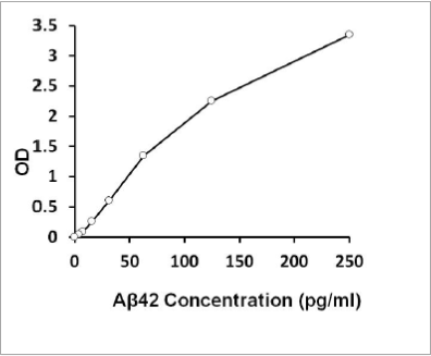
Sensitivity: 2 pg/ml
Dynamic Range: 3.9-250 pg/mL
Incubation Time: 1 hour
Sample Type: Cell and tissue lysate as well as in body fluids
Sample Size: 100 µL
Alternative Names: Aβ42, Mouse / rat beta-Amyloid (1-42)


SAMPLE COLLECTION & STORAGE INFORMATION
The sample collection and storage conditions listed below are intended as general guidelines. Sample stability has not been evaluated.
Note: protease inhibitor cocktail with PMSF must be added to all samples to avoid protein degradation.
Brain lysate- Mix 800 μl of homogenate buffer (5M Guanidine HCl, 50 mM Tris HCl pH=8.0) with 100 mg of brain sample in a Dounce homogenizer placed on ice. Homogenize the tissue thoroughly and incubate at room temperature for 3-4 hours with mixing. Guanidine-HCl brain homogenates are stable at 4°C for several weeks and can be freeze-thawed many times without degradation of beta-Amyloid peptides.

 
Plasma – Collect plasma using EDTA or heparin as an anticoagulant. Centrifuge for 15 minutes at 1000 x g within 30 minutes of collection. Assay immediately or aliquot and store samples at ≤ -20 °C. Avoid repeated freeze-thaw cycles.


Assay Background

Alzheimer’s Disease (AD) is the most common neurodegenerative disorder in elderly people. It has been demonstrated that AD has biological causes and is characterized by the presence of senile plaques and neurofibrillary tangles mainly in cerebral cortex and hippocampus brain regions. Beta-Amyloid (1-40) (Aβ40) and beta-Amyloid (1-42) (Aβ42) are the main components of the above plaques; however, other forms of beta-Amyloid peptides are also present. Both peptides are cleaved from the Amyloid Precursor Protein (APP) by β-secretase and γ-secretase enzymes. Many studies suggest that Aβ42 or/and Aβ43 are required to initiate formation of amyloid plaques and neurofibrills that leads to the neurodegeneration, while Aβ40 is less neurotoxic.


Related Products

Mouse/Rat beta-Amyloid (1-42) ELISA Assay Kit (plasma)
Mouse/Rat beta-Amyloid (1-40) ELISA Assay Kit
beta-Amyloid (1-42) ELISA Assay Kit

Additional Information

Assay Principle


This assay employs the quantitative sandwich enzyme immunoassay technique. A monoclonal antibody specific for A 42 has been pre-coated onto a microtiter plate. Standards or samples are pipetted into the wells together with HRP-labeled A 42 antibody. After incubation with continuous agitation, the capture antibody-antigen-detection antibody complex is developed and immobilized to the pre-coated wells. After washing away any unbound reagents, a substrate solution (TMB) is added to the wells and color develops in proportion to the amount of A 42 bound in the initial step. The color development is stopped by the addition of acid and the intensity of the color is measured at a wavelength of 450nm. The concentration of A 42 in the sample is then determined by comparing the O.D of samples to the standard curve. The concentration of antigen is directly proportional to the optical density measured in the wells.

Assay Procedure


  1. Remove excess microplate strips from the plate frame, return them to the foil pouch containing the desiccant pack, and reseal it.
  2. Add 100 μl of standards in duplicates including blank and 100 μl of diluted samples into wells.
  3. Add 50 μl of diluted HRP-labeled detection antibody into each well, cover the plate and incubate the plate overnight at 4°C in dark.
  4. Aspirate each well and wash, repeating the process 6 times for a total 7 washes. Wash by filling each well with 1× Wash Buffer (350 μl) using a squirt bottle, manifold dispenser, or autowasher. Keep the wash buffer in the wells for 20 seconds before remove it. Complete removal of liquid at each wash step is essential to good performance. After the last wash, remove any remaining Wash Buffer by aspirating, decanting or blotting against clean paper towels.
  5. Add 100 μl of TMB Reagent to each well and tap the plate gently to mix the reagent. Incubate for 5-15 minutes at room temperature in dark.
  6. Add 50 μl of Stop Solution to each well and tap the plate gently to mix the reagent. The color of the solution should change from blue to yellow.
  7. Read the OD with a microplate reader at 450nm immediately. It is recommended reading the plate within 20 min after adding the stop solution.

Typical Standard Curve


Package Inserts


Please note: All documents above are for reference use only and should not be used in place of the documents included with this physical product. If digital copies are needed, please contact us.