HMGB1 Serum ELISA Assay

$1,635.00

The Eagle Biosciences HMGB1 ELISA Assay kit is intended for the quantification of Human / Mouse / Rat HMGB1 in serum samples. The Eagle Biosciences HMGB1 ELISA assay kit is for research use only and should not be used for diagnostic procedures.

SKU: ARG81351 Categories: , ,

HMGB1 Serum ELISA Assay

The HMGB1 Serum ELISA Assay is For Research Use Only

Size: 1 x 96 wells
Sensitivity: 0.4 pg/ml
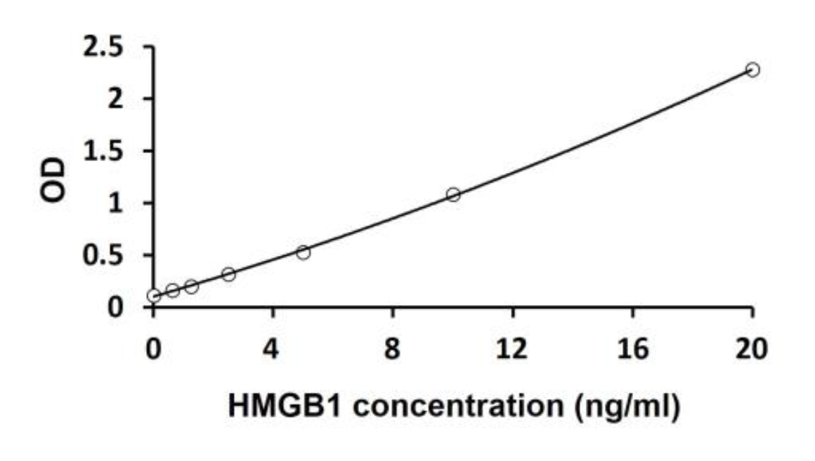
Dynamic Range: 0.625-20 ng/ml
Incubation Time: 2 hours
Sample Type: Serum
Sample Size: 50 µl
Alternative Names: Multispecies High Mobility Group Box 1


Assay Principle

This assay employs the sandwich enzyme immunoassay technique for the detection of Human/Mouse/Rat HMGB1 in serum samples. An antibody specific for HMGB1 has been pre-coated onto a microtiter plate. Standards or samples are pipetted into the wells and any HMGB1 present is bound on the plate. After washing away any unbound substances, a Horseradish Peroxidase (HRP) conjugated primary antibody binds to HMGB1 is added to each well and incubates. After washing away any unbound antibody-enzyme reagent, a substrate solution (TMB) is added to the wells and color develops in proportion to the amount of total HMGB1 bound in the initial step. The color development is stopped by the addition of acid and the intensity of the color is measured at a wavelength of 450nm ±2nm. The concentration of total HMGB1 in the sample is then determined by comparing the O.D of samples to the standard curve.


Related Products

HMGB1 Plasma ELISA Assay
Rodent HMGB1 ELISA Assay
HDGF ELISA Assay Kit

Additional Information

Assay Background


Multifunctional redox sensitive protein with various roles in different cellular compartments. In the nucleus is one of the major chromatin-associated non-histone proteins and acts as a DNA chaperone involved in replication, transcription, chromatin remodeling, V(D)J recombination, DNA repair and genome stability. Proposed to be a universal biosensor for nucleic acids. Promotes host inflammatory response to sterile and infectious signals and is involved in the coordination and integration of innate and adaptive immune responses. In the cytoplasm functions as sensor and/or chaperone for immunogenic nucleic acids implicating the activation of TLR9-mediated immune responses, and mediates autophagy. Acts as danger associated molecular pattern (DAMP) molecule that amplifies immune responses during tissue injury. Released to the extracellular environment can bind DNA, nucleosomes, IL-1 beta, CXCL12, AGER isoform 2/sRAGE, lipopolysaccharide (LPS) and lipoteichoic acid (LTA), and activates cells through engagement of multiple surface receptors. In the extracellular compartment fully reduced HMGB1 (released by necrosis) acts as a chemokine, disulfide HMGB1 (actively secreted) as a cytokine, and sulfonyl HMGB1 (released from apoptotic cells) promotes immunological tolerance.

Assay Procedure


  1. Remove excess microplate strips from the plate frame, return them to the foil pouch containing the desiccant pack, and reseal it.
  2. Add 50 µl of standards, samples and zero controls into appropriate wells.
  3. Add 50 µl of Assay buffer into all wells immediately. Mix thoroughly by gently shaking or tapping the plate. Cover the plate and incubate for overnight (~16 hours) at 4°C.
  4. Aspirate each well and wash, repeating the process 2 times for a total 3 washes. Wash by filling each well with 1X cold wash buffer (350 μl) using a squirt bottle, manifold dispenser, or autowasher. Complete removal of liquid at each is essential to good performance. After the last wash, remove any remaining buffer by aspirating, decanting or blotting against clean paper towels.
  5. Add 100 µl 1X HRP-antibody conjugate into each well. Mix thoroughly by gently shaking or tapping the plate. Cover wells and incubate for 1 hour at 37°C.
  6. Aspirate each well and wash as step 4.
  7. Add 100 μl of TMB substrate to each well. Incubate for 10 minutes at RT in dark. Substrate will change from colorless to different strengths of blue.
  8. Add 50 μl of Stop solution to each well. The color of the solution should change from blue to yellow. Mix thoroughly by gently shaking the plate.
  9. Read the OD with a microplate reader at 450 nm immediately.

Standard Curve


HMGB1 Serum ELISA Assay

Package Inserts


Please note: All documents above are for reference use only and should not be used in place of the documents included with this physical product. If digital copies are needed, please contact us.

Product Citations