High Sensitive Anti-TPO ELISA

$360.00

The Anti-TPO (Thyroperoxidase) ELISA Assay Kit is intended for the quantitative determination of Anti-TPO in serum by enzyme linked immunoassay (ELISA). The Eagle Biosciences Anti-TPO (Thyroperoxidase) ELISA Assay kit is for research use only and not to be used in diagnostic procedures.

SKU: TPO31-K01 Categories: , ,

High Sensitive Anti-TPO ELISA

High Sensitive Anti-TPO ELISA Developed and Manufactured in the USA

Size: 1×96 wells
Sensitivity: 1 U/mL
Dynamic Range: 15 – 960 U/ml
Incubation Time: 2 hours
Sample Type: Serum
Sample Size: 10 µL
Alternative Names: Anti-TPO IgG, Thyroperoxidase IgG
For Research Use Only

Controls Included


Assay Principle

The High Sensitive Anti-TPO ELISA is designed, developed and produced for the quantitative measurement of human anti-TPO IgG level in test sample. The assay utilizes the streptavidin coated microplate based enzyme immunoassay technique.

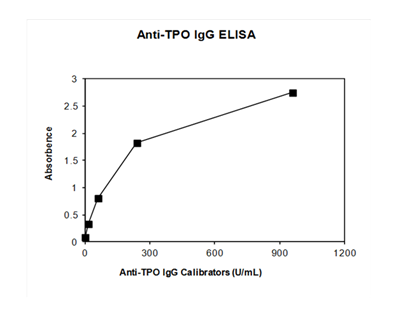
Assay calibrators, controls and pre-diluted human serum samples containing anti-TPO IgG are added to microtiter wells of microplate that was coated with high affinity streptavidin on its wall. The autoantibody reaction would not start until the addition of a biotinylated human TPO antigen. After the first incubation period, the unbound protein matrix was removed in the subsequent washing step. A horseradish peroxidase-conjugated rabbit anti-human IgG subclass specific antibody (tracer antibody) is added to each well. After an incubation period an immunocomplex of “solid-phase bound biotin-TPO – human anti-TPO IgG – HRP-conjugated tracer antibody” is formed if there is human anti-TPO IgG autoantibody present in the test sample. The unbound tracer antibody is removed in the subsequent washing step. HRP-conjugated tracer antibody bound to the well is then incubated with a substrate solution in a timed reaction and then measured in a spectrophotometric microplate reader. The enzymatic activity of the tracer antibody bound to the human IgG on the wall of the microtiter well is directly proportional to the amount of human anti-TPO IgG autoantibody level in the sample. Plotting the absorbance versus the respective human anti-TPO IgG autoantibody concentration for each calibrator on point-to-point or 4-parameter fit generates a calibrator curve. The concentration of human anti-TPO IgG autoantibody in test samples is determined directly from this calibrator curve.


Related Products

Mouse Thrombopoietin ELISA Kit
High Sensitive Anti-Tg ELISA

Additional Information

Assay Background


It is a routine practice of measuring serum autoantibodies to thyroglobulin (Tg) and microsomal (TPO) for aid in detecting and monitoring autoimmune thyroid disease. Serum anti-TPO autoantibody and anti-Tg autoantibody are found to be well correlating with histological changes in Harshimoto’s thyroiditis. Clinically, positive anti-TPO autoantibody is detected in patients with chronic thyroiditis (70-90%), primary hypothyroidism (~60%), thyrotoxicosis (~50%) and thyroid tumors (~17%), however, anti-Tg autoantibody is mainly identified in patients with Harshimoto’s thyroiditis and Graves’ disease (40-70%).

Although ELISA technology has applied to detecting these autoantibodies, the high background in normal population would decrease the clinical diagnostic sensitivity and specificity. This high sensitive anti-TPO autoantibody IgG ELISA Assay Kit was developed with proprietary technology that leads to a very low reaction background in normal population and thus would increase the clinical diagnostic sensitivity and specificity.

Assay Procedure


  1. Add 25 µL of calibrators, controls and diluted patient serum samples into the designated microwell.
  2. Add 100 µL of biotinylated TPO solution into each well.
  3. Cover the plate with one plate sealer.
  4. Incubate plate at room temperature for 1 hour.
  5. Prepare Anti-hIgG Tracer Antibody Working Solution by 1:21 fold dilution of the tracer antibody with the Tracer Antibody Diluent. For each strip, it is required to mix 1 mL of Tracer Antibody Diluent with 50 µL of the Tracer Antibody in a clean test tube.
  6. Remove the plate sealer. Aspirate the contents of each well. Wash each well 5 times by dispensing 350 µL to 400 µL of working wash solution into each well and then completely aspirating the contents. Alternatively, an automated microplate washer can be used.
  7. Add 100 µL of above diluted tracer antibody working solution to each of the wells.
  8. Cover the plate with one plate sealer and also with aluminum foil to avoid exposure to light.
  9. Incubate plate at room temperature for 30 minutes.
  10. Remove the plate sealer. Aspirate the contents of each well. Wash each well 5 times by dispensing 350 µL to 400 µL of working wash solution into each well and then completely aspirating the contents. Alternatively, an automated microplate washer can be used.
  11. Add 100 µL of ELISA HRP Substrate into each of the wells.
  12. Cover the plate with a new plate sealer and also with aluminum foil to avoid exposure to light.
  13. Incubate plate at room temperature for 20 minutes.
  14. Remove the aluminum foil and plate sealer. Add 100 µL of ELISA Stop Solution into each of the wells. Mix gently.
  15. Read the absorbance at 450 nm within 10 minutes in a microplate reader.

Typical Standard Curve


High Sensitive Anti-TPO ELISA

Package Inserts


Please note: All documents above are for reference use only and should not be used in place of the documents included with this physical product. If digital copies are needed, please contact us.

Product Citations

Confirmation and Identification of Biomarkers Implicating Environmental Triggers in the Pathogenesis of Type 1 Diabetes
Harms, Ostlund, Cabrera et al
Front Immunol (2020) 11, 1922 DOI: 10.3389/fimmu.2020.01922
Snippet: IgG(88-50550), total IgM (88-50620), IL-18 (BMS267), high sensitivity IL-17A (BMS2017HS), high sensitivity TNF-α (BMS223HS), high sensitivity IL-10(BMS215HS), and high sensitivity IFN-γ (BMS228HS);from AbCam MDC (ab223866);from Eagle Biosciences GAD65
Showing 1-1 of 1 papers.
Powered by BizGenius