Gliadin IgA ELISA Kit

$435.00

The GliaDea IgA ELISA Assay Kit is used for the quantitative determination of IgA antibodies against deamidated gliadin in human serum or plasma for the diagnosis of celiac disease. The Eagle Biosciences Gliadin IgA ELISA Kit is for research use only.
This product was previously known as GDA31-K01.

Gliadin IgA ELISA Kit

Gliadin IgA ELISA Kit Developed and Manufactured by Medipan

Size: 1×96 wells
Sensitivity: 1 U/ml
Dynamic Range: 1 – 300 U/ml
Incubation Time: 2 hours
Sample Type: Serum, Plasma
Sample Size: 10 µL
Alternative Names: GliaDea IgA ELISA, Human Gliadin IgA ELISA
For Research Use Only

Controls Included


Reference Values
Quantitative (U/ml): Negative < 10, Positive > 15, Grey Zone 10-15
Semi-quant. BI: Negative < 1.0, Positive > 1.5, Grey Zone 1.0-1.5

Specimens with concentrations detected in the grey zone should be tested again.
It is recommended that each laboratory establishes its own normal and pathological reference ranges for serum anti-deamidated gliadin IgA levels, as usually done for other diagnostic parameters, too. Therefore, the above mentioned reference values only provide a guide to values which might be expected.

Assay Background

Celiac disease (CD, sprue, gluten enteropathy) is an intolerance to gluten, a cereal protein present primarily in wheat. This gluten-induced enteropathy leads to a severe damage of the small intestine and a so-called “flat” mucosa. Due to this extensive lesions mal-absorption occurs accompanied with a depletion of key nutrients.

Gliadin the alcohol soluble fraction of gluten represents the causative agent of celiac disease. During resorption gliadin is deamidated by tissue transglutaminas (tTG). Linked to specific genetic predisposition parts of this deamidated gliadin activate HLA-DQ2 and -DQ8 antigen presenting cells. These provoke an inflammatory process in the small intestine triggering both cellular and humoral immune responses. Hence pathogenic Tissue damage as well as production of antibodies against deamidated gliadin and tTG occurs.

Celiac disease is one of the most common enteropathies found in infants, showing first symptoms already at the age between sixth and eighteen months. Incidence rates for celiac disease range from 1 in 300 (Western Ireland) to 1 in 4700 in European countries. However, a high number of subclinical cases of celiac disease have been detected by in-vitro tests revealing a prevalence of 4 in 1000. Individuals suffering from prolonged celiac disease additionally face an elevated risk of developing T cell lymphoma.


Related Products

Anti-Gliadin IgG ELISA Kit
Anti-Gliadin sIgA / IgA ELISA Assay Kit
Celiac EmA (Anti-Endomysium Antibody) IgA ELISA Assay Kit

Additional Information

Assay Principle


The antibodies of the calibrators, positive control, and diluted patient samples react with fragments of deamidated gliadin immobilized on the solid phase of microtiter plates. After an incubation period of 60 min at room temperature (18…25°C), unbound serum components are removed by a wash step.

The bound autoantibodies react specifically with anti-human-IgA-antibodies conjugated to horseradish peroxidase (HRP) within the incubation period of 30 min at room temperature. Excessive conjugate is separated from the solid-phase immune complexes by the following wash step.

HRP converts the colorless substrate solution of 3,3’,5,5’-tetramethyl¬benzidine (TMB) added into a blue product. This enzyme reaction is stopped by dispensing an acidic solution (H2SO4) into the wells after 15 min at room temperature turning the solution from blue to yellow.

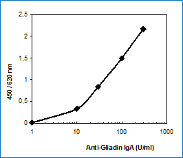
The optical density (OD) of the solution at 450 nm is directly proportional to the amount of specific antibodies bound. The standard curve is established by plotting the concentrations of the antibodies of the calibrators (x-axis) and their corresponding OD values (y-axis) measured. The concentration of antibodies of the specimen is directly read off the standard curve. Evaluating the test by a semi-quantitative method is also possible.

Assay Procedure


  1. Bring all reagents to room temperature (18…25°C) before use. Mix gently, avoid foam.
  2. Dispense 100 µl calibrators (0 optional) 1 – 4 (quantitative) or 100 µl calibrator 1 (semi-quantitative)100 µl control P (N optional) 100 µl diluted patient samples into the respective wells.
  3. Seal plate, incubate 60 min at room temperature.
  4. Decant, then wash each well three times using 300 µl wash solution (made of B).
  5. Add 100 µl of conjugate (D) solution to each well.
  6. Seal plate, incubate 30 min at room temperature.
  7. Decant, then wash each well three times using 300 µl wash solution (made of B).
  8. Add 100 µl of substrate (E) to each well.
  9. Incubate 15 min protected from light at room temperature.
  10. Add 100 µl of stop solution (F) to each well and mix gently.
  11. Read the OD at 450 nm versus 620 or 690 nm within 30 min after adding the stop solution.

Typical Standard Curve


Package Inserts


Please note: All documents above are for reference use only and should not be used in place of the documents included with this physical product. If digital copies are needed, please contact us.

Product Citations