Anti-GP2 IgG ELISA Kit

$435.00

The Anti-GP2 IgG is used for the quantitative determination of IgG antibodies against glycoprotein 2 (GP2) in human serum for the diagnosis of Crohn’s disease. The Eagle Biosciences Anti-GP2 IgG is for research use only and not for use in diagnostic procedures.
This product was previously known as G2G31-K01.

Anti-GP2 IgG ELISA Kit

Anti-GP2 IgG ELISA Kit Developed and Manufactured by Medipan

Size: 1×96 wells
Sensitivity: 1 U/ml
Dynamic Range: 1 – 300 U/ml
Incubation Time: 1.5 hours
Sample Type: Serum, Plasma
Sample Size: 10 µL
Alternative Names: Glycoprotein 2
For Research Use Only


Assay Principle

The Anti-GP2 IgG ELISA Assay Kit is an enzyme immunoassay for the quantitative determination of IgG antibodies to glycoprotein 2.

The antibodies of the calibrators, positive control, and diluted patient samples react with the antigen immobilized on the solid phase of microtiter plates. After an incubation period of 60 min at room temperature (18…25°C), unbound serum components are removed by a wash step. The bound IgG autoantibodies react specifically with anti-human-IgG-antibodies conjugated to horseradish peroxidase (HRP) within the incubation period of 30 min at room temperature. Excessive conjugate is separated from the solid-phase immune complexes by the following wash step.

HRP converts the colorless substrate solution of 3,3’,5,5’-tetramethyl¬benzidine (TMB) added into a blue product. This enzyme reaction is stopped by dispensing an acidic solution (H2SO4) into the wells after 15 min at room temperature turning the solution from blue to yellow.

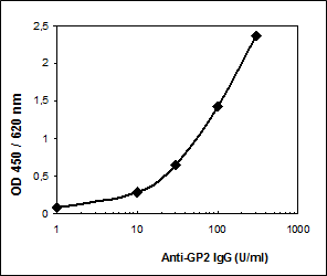
The optical density (OD) of the solution at 450 nm is directly proportional to the amount of specific antibodies bound. The standard curve is established by plotting the concentrations of the antibodies of the calibrators (x-axis) and their corresponding OD values (y-axis) measured. The concentration of antibodies of the specimen is directly read off the standard curve.


Related Products

Anti-GP2 IgA ELISA Kit
Anti-Beta 2 GP-1 (Glycoprotein 1) ELISA Assay Kit
Anti-GP2 (Glycoprotein 2) IgG ELISA Assay Kit

Additional Information

Assay Background


Non-specific inflammatory bowel disease including Crohn’s disease (Enteritis regionalis) and ulcerative colitis (UC) are characterised by unknown etiology as well as chronic-remitting inflammatory processes of the intestine. Whereas the inflammation of ulcerative colitis is restricted to the mucosa and submucosa of colon and rectum, Crohn’s disease (CD) shows a wide spread inflammation of gastro-intestinal tract with granuloma formation. The risk developing one of these diseases is strongly correlated to immunologic, genetic, infectious and environmental factors.

The differential diagnosis of inflammatory bowel diseases to chronic diarrhea, recurrent abdominal dolor, infectious colitis, anorexia as well as the differentiation of CD to UC is still a challenge.

Autoantibodies of exocrine pancreas (PAB) were identified as specific serological marker for CD. A prevalence of 39 % of these autoantibodies in patients with CD could be demonstrated by indirect immune fluorescence (Stöcker et al. 1987). Glycoprotein 2 (GP2), a membrane-bound pancreatic protein, could be identified/verified as the major target of PAB’s (Roggenbuck et al., 2009). In combination with the detection of autoantibodies to Saccharomyces cerevisiae (ASCA) with a prevalence of 70 % in patients with CD and atypical antineutrophile cytoplasmatic antigens (aANCA) which are mainly found in patients with UC, PAB’s against GP2 could be used as a highly specific serological marker for differential diagnosis of CD to UC.

Assay Procedure


  1. Bring all reagents to room temperature (18…25°C) before use. Mix gently, avoid foam.
  2. Dispense 100 µl calibrators (0 optional) 1 – 4 100 µl control P (N optional) 100 µl diluted patient samples into the respective wells.
  3. Seal plate, incubate 60 min at room temperature.
  4. Decant, then wash each well three times using 300 µl wash solution (made of B).
  5. Add 100 µl of conjugate (D) solution to each well.
  6. Seal plate, incubate 30 min at room temperature.
  7. Decant, then wash each well three times using 300 µl wash solution (made of B).
  8. Add 100 µl of substrate (E) to each well.
  9. Incubate 15 min protected from light at room temperature.
  10. Add 100 µl of stop solution (F) to each well and mix gently.
  11. Read the OD at 450 nm versus 620 or 690 nm within 30 min after adding the stop solution.

Typical Standard Curve


Package Inserts


Please note: All documents above are for reference use only and should not be used in place of the documents included with this physical product. If digital copies are needed, please contact us.

Product Citations