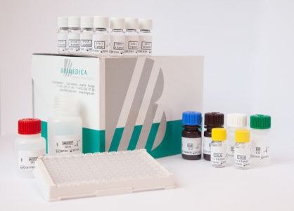
VEGF ELISA Assay Kit

$545.00

The VEGF ELISA Assay Kit is intended for the quantitative determination of human VEGF in serum, EDTA plasma, and citrate plasma. The Eagle Biosciences VEGF ELISA is for research use only and not for use in diagnostic or clinical procedures.

VEGF ELISA Assay Kit

VEGF ELISA Assay Kit Developed and Manufactured in Austria by Biomedica

Size: 1×96 wells
Sensitivity: 2.5 pg/ml
Standard Range: 0 – 2000 pg/ml
Incubation Time: 4.5 hours
Sample Type: Serum, EDTA plasma, citrate plasma, cell culture supernatants, urine
Sample Size: 10 µl
Alternative Names: Vascular endothelial growth factor, VEGF-A, VEGFA, VPF, MVCD1, Vascular endothelial growth factor A, vascular endothelial growth factor A121, vascular endothelial growth factor A165, Vascular permeability factor, VEGF-A, VPF
For Research Use Only

Controls Included


Assay Principle

The human Vascular Endothelial Growth Factor (VEGF) ELISA kit is a sandwich enzyme immunoassay that has been optimized and fully validated for the quantitative determination of human VEGF in serum, EDTA plasma, and citrate plasma. Validation experiments have been performed according to international quality guidelines (ICH/ FDA/ EMEA). Cell culture supernatant and urine samples are compatible with this ELISA. The VEGF ELISA assay recognizes both natural and recombinant human VEGF. The assay employs highly purified epitope mapped antibodies as well as human serum-based standards and controls.
In a first step, assay buffer is pipetted into the wells of the microtiter strips. Thereafter, STD/ sample/CTRL are pipetted into the wells, which are pre-coated with the recombinant antihuman VEGF antibody. Any soluble VEGF present in the STD/sample/CTRL binds to the pre-coated anti-VEGF antibody in the well. After incubation, a washing step is applied where all non-specific unbound material is removed. In the next step, the biotinylated anti-VEGF antibody (AB) is pipetted into the wells and reacts with the VEGF present in the sample, forming a sandwich.
Next, all unbound antibody is removed during another washing step. In the following step, the conjugate (streptavidin-HRPO) is added and reacts with the biotinylated anti-VEGF antibody. After another washing step, the substrate (tetramethylbenzidine; TMB) is pipetted into the wells. The enzyme catalysed color change of the substrate is directly proportional to the amount of VEGF present in the sample. This color change is detectable with a standard microtiter plate ELISA reader.
A dose response curve of the absorbance (optical density, OD at 450 nm) versus standard concentration is generated, using the values obtained from the standards. The concentration of soluble VEGF in the sample is determined directly from the dose response curve.

Related Products

Mouse VEGF ELISA Assay
Human sVEGFR1 ELISA Kit
Human VEGFR2/KDR ELISA Kit

Additional Information

Assay Background


Vascular endothelial growth factor (VEGF or VEGF-A), is a growth hormone secreted by endothelial cells, fi broblasts, smooth muscle cells, platelets, macrophages, and many other cell types (https://ebi16.uniprot.org/uniprot/P15692). It belongs to the cysteine-knot growth factor superfamily (1) and has a molecular weight of about 40 kDa. Currently, 17 different VEGF isoforms have been described to be expressed from one single gene. They are produced by alternative promoter usage/initiation or alternative splicing/proteolysis after protein translation. The N-terminal region is responsible for receptor binding and conserved among all VEGF isoforms. In contrast, residues of the C-terminus differ between isoforms and determine protein length and properties: binding to co-receptor Neuropilin-1 (NRP1) or to extracellular matrix (ECM), agonist/antagonist of angiogenesis. Most isoforms result from the common transcripts: VEGF111, VEGF121, VEGF145, VEGF165, VEGF189 and VEGF206. Additionally, a third VEGF variant (VEGFAx), that demonstrates pro- and anti-apoptotic properties, was described. Thus, vascularization is tightly controlled by the balance of various splice variants, their availability and concentration, whereas isoforms linked to the ECM constitute a reservoir of VEGF that can quickly be shed to circulating forms (1). One of the most potent pro-angiogenic isoforms is VEGF165a. After secretion, 50-70% of VEGF165a is attached to the extracellular matrix (via heparin binding site), the rest is freely diffusible (1) . It is the most abundant isoform and enhances signaling over the VEGFR2 receptor by additionally binding to its co-receptor Neuropilin-1. VEGF A isoforms are glycosylated, homodimeric proteins. Two anti-parallel monomers are linked by intermolecular disulfide bonds (2) whereas eight cysteine residues form a knot-like structure at one end of each monomer (3). However, heterodimerization with PLGF has been described as well (4).

Typical Standard Curve


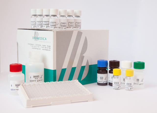
VEGF ELISA Assay Kit

Package Inserts


Please note: All documents above are for reference use only and should not be used in place of the documents included with this physical product. If digital copies are needed, please contact us.

Product Citations