RF IgA ELISA Kit
RF IgA ELISA Kit Developed and Manufactured by Medipanm>
Size: 1×96 wells
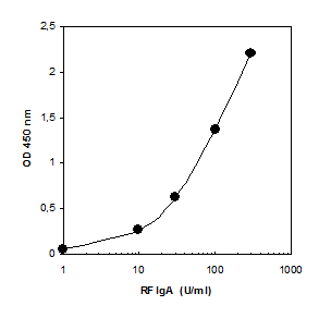
Sensitivity: 1 U/ml
Dynamic Range: 1 – 300 U/ml
Incubation Time: 2.5 hours
Sample Type: Serum, Plasma
Sample Size: 10 µL
Alternative Names: Rheumatoid Factor IgA ELISA, Human RF IgA ELISA, Human Rheumatoid Factor IgA ELISA
For Research Use Only
Scientific Background
Patients suffering from rheumatoid arthritis (RA) exhibit RF autoantibodies recognizing the Fc part of IgG. RA or chronic polyarthritis has a yet unknown etiology and represents the most frequent rheumatic inflammatory disorder demonstrating a prevalence of up to 1%. One of the typical manifestations of RA is symmetric synovialitis of limb joints often accompanied by involvement of the cervical spinal column.
Beside clinical features one of the criteria of the American College of Rheumatology for the classification of RA is the presence of RF (1). Up to 80 % of RA patients may demonstrate RF. RF can occur years prior to the onset of disease and RF positive apparently healthy individuals bear a 5 – 40 times higher risk to develop RA (2). However, patients suffering from other autoimmune, infectious or B-cell lympho¬pro-liferative disorders as well as apparently healthy elderly individuals may develop RF.
High concentrations of RF are often associated with a more severe disease comprising a faster destruction of joints. In addition, they are found in patients with extra-articular manifestations such as rheumatoid nodules, polyneuropathy, vasculitis or Sicca syndrome.
RF may belong to the IgG, IgM or IgA isotype whereas IgM RF is the most frequent isotype to be determined in RA patients. Extra-articular manifestations seem to be associated with IgA RF. Like RF of the IgM isotype high concentrations of IgG RF seems to appear with patients suffering from more progressive erosions of joints. In long- time RA IgA and IgG RF are considered to be prognostic markers for systemic manifestation.
Products Related to RF IgA ELISA Kit
RF IgM (Rheumatoid Factor) ELISA Assay Kit
RF IgG (Rheumatoid Factor) ELISA Assay Kit
Celiac EmA (Anti-Endomysium Antibody) IgA ELISA Assay Kit