What
are Endothelins?
Endothelins are a family of peptides, which comprises endothelin-1
(ET-1), endothelin-2 (ET-2) and endothelin-3 (ET-3), each containing 21
amino-acids.Endothelins were
discovered in 1988 when isolated from porcine aortic endothelial cells. They are long-acting vasoconstrictor substances
that play a strong, important role in blood pressure regulation and sodium and
fluid homeostasis. Other functions of
the ET system involve renal microcirculation regulation and they have been
found to contribute to the pathogenesis of renal injury.
The biological role of ET extends
beyond regulating vascular tone also in its growth regulatory properties. The
peptide interacts in an autocrine/paracrine manner with specific ET receptors
found on numerous cells, including smooth muscle cells, myocytes, and
fibroblasts. The half-life of ET in the
plasma is less than one minute, whereas clearance of Big ET is much slower,
therefore Big ET more accurately reflects the relative physiological effect.
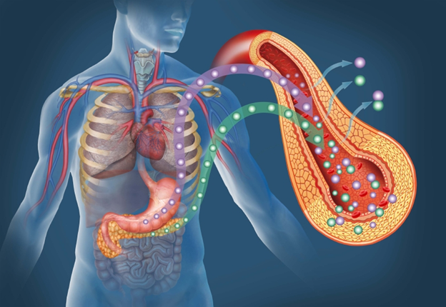
Endothelin has been identified in a variety of tissues, including lung, kidney,
brain, pituitary and peripheral endocrine tissues and placenta.
Summary of Biological Functions:
Regulation of:
- Blood pressure
- Vascular tone
- Sodium and fluid homeostasis
- Renal microcirculation
- Autocrine and paracrine functions
Why Measure Endothelins?
Since the principal actions of
endothelin surround increasing blood pressure and vascular tone, endothelin
antagonists may serve to play important parts in offsetting the negative impact
endothelin has in invivo. Past studies have focused on observing the mediation
of the pathogenesis of hypertension and its complications. More specifically,
some studies have demonstrated that ET-1 plays an significant role in the
progression of coronary artery disease(CAD) and subsequent plaque rupture and
ACS onset. In addition, findings from other studies have proven that Big ET-1
and ET-1 are strong independent predictors of survival in patients with severe
CHF. Based on this research, it is
believed that anti-endothelin therapy can help to improve symtoms and
potentially be used as treatment in such conditions as vascular remodelling,
left ventricular hypertrophy, hypertensive kidney damage and atherosclerosis. However, due to the complex nature of the
endothelin system, further research may be necessary to further define these
biological intricacies for the development of therapeutic agents. Robust, sensitive, and specific immunoassays
could be valuable tools to assist researchers in these investigations.
Biosynthesis
of Endothelins
The synthetic Endothelin pathway begins with a 212
amino acid peptide, called preproendothelin-1 which is then converted to
proendothelin-1 following a cleavage and removal of a short secretory
sequence. Furin (A protein that in
humans is encoded by the FURIN gene, its main function is to cleave proteins
just downstream of a basic amino acid target sequence) then prompts the
cleavage of proendothelin-1 to create biologically-inactive, 38 amino-acid
precursor called Big Endothelin. Big Endothelin-1
is then cleaved to yield endothelin-1 prompted by the action of several
endothelin-converting enzymes (ECE). Big Endothelin-1 can also be hydrolyzed by
chymase (serine proteases that have chymotrypsin-like cleavage properties) to
generate endothelin (1-21) in vitro.
What is Big Endothelin 1?
Big Endothelin-1 is a biologically-inactive
peptide and 38 amino-acid precursor with a plasma half-life of 30 minutes. Since
Plasma ET-1 has a much shorter half-life (about 1.5 min) and it has proven to
be problematic to measure circulating concentrations, Big Endothelin-1 has
become an excellent tool for measuring and monitoring the endothelin system
activation due to its inherent stability.
What is Endothelin 1-21?
Endothelin 1-21 is a potent vasoconstrictor and is created by the cleavage of Big Endothelin by a membrane-bound metalloproteinase, the Endothelin Converting Enzyme (ECE). This process yields active ET 1-21 and also the biological inactive C-terminal fragment (22-38) as described above.
Research and Indications:
The intrinsic biological function of Big ET-1 is
still under investigation as many aspects of its role are still unknown. However, recent studies suggest that this
analyte may work in opposition to ET-1 utilizing ET receptors and thereby
stimulating diueresis and natriuresis.
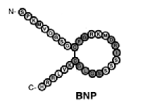
In fact, a very large study, Masson et al 2006 revealed
that ET stimulates the release of natriuretic peptides: ET participates in the
mechanical stretch-induced release of BNP by atrial myocytes. This research involved monitoring and
measuring various analytes with Big ET-1 serving as the main focus of study for
the cohort of 2,300 heart failure patients. This research also clearly
demonstrated that plasma concentrations of BNP show a strong and independent
association with Big ET using our Big Endothelin-1 ELISA Assay Kit.
In past studies, it has been
discovered that Big-Endothelin levels raise significantly in patients with
various types of tumors and increased concentrations of this analyte have been
corrleated to worse outcomes. Teng et al 2006 investigated the levels of Big-Endothelin-1 in plasma of
gastric carinoma patients before and after radical gastroectomy utilizing our Big Endothelin-1 ELISA Assay Kit. It was evident from the results of this study
that there is a corrleation between the increase in Big ET-1 plasma levels and
the progression of tumors. Therefore, not
only could this analyte serve as a tumor marker for gastric cancer but it could
also serve as a powerful tool in monitoring recurrent disease.
Another
interesting study Arun et. al 2004 that utilized our Big Endothelin-1 ELISA Assay Kit, evaluated
the expression of Big ET-1 and ET-1 in non-small cell lung cancer (NSCLC). This was the first study to investigate this
analyte in this particular area of cancer research and more specifically, NSCLC. Since lung cancer is one of the most common
causes of cancer-related death in the Western world, there has been a great
need to further explore and understand the molecular biology of this
diseases. It discovered from the
results of this study that ET-1 and Big ET-1 are synthesized by NSCLC tumor in
vitro as well as in vivo and elevated levels have been associated with poor
outcomes. This evidenece has lead
invesigators to believe that ET-1 and Big ET-1 could serve as strong biomarkers
of this disease and as potential novel targets for treatment of NSCLC.
References:
- Agapitov, Alexei et al. “Role of Endothelin in Cardiovascular Disease.” JRAAS, 2002; 3:1-15.
- Arun, C. et al. Endothelin-1 is a Novel Prognostic Factor in Non-Small Cell Lung Cancer.” Journal of Biological Markers 2004; 19 (4):262-267.
- Beneden, Van et al. “Superiority of big endothelin-1 and endothelin-1 over natriuretic peptides in predicting survival in severe congestive heart failure: a 7-year follow-up study.”J Card Fail. 2004 Dec; 10(6):490-5.
- Burg MM et al. “Depression Predicts Elevated Endothelin-1 in Patients With Coronary Artery Disease.” Psychosom Med 2011; 73: 2-6.
- Harrison-Benard, Lisa et al. “Enhanced Vascular Chymase-Dependent Conversion of Endothelin in the Diabetic Kidney.” The Oschner Journal 2013 13:1, 49-55.
- Jiao, Wenjie et al. “Elevation of circulating big endothelin-1: an independent prognostic factor for tumor recurrence and survival in patients with esophageal squamous cell carcinoma.” BMA Cancer 2008, 8:334.
- Masson et al. “The Prognostic Value of Big Endothelin-1 in More than 2,300 Patients with Heart Failure Enrolled in the Valsartan Heart Failure Trial (Val-HeFT).”Journal of Cardiac Failure 2006; 12: 375-380.
- Teng et al. “Pre- and Post-operative Plasma Big-Endothelin-1 Levels in Patients with Gastric Carcinoma Undergoing Radical Gastrectomy.” Anticancer Research 2006; 26: 2503-2508.
- Verghese, Mathew et al. “Clinical Implications of a Sandwich Enzyme Immunoassay for Big Endothelin-1.” Clinical Chemistry 1997; 43: 9-10.
Related Kits:
Endothelin (1-21) ELISA Assay Kit
Big Endothelin-1 ELISA Assay Kit
Cardiovascular Assay Kits
Related News:
Eagle Biosciences Introduces Endothelin 1-21 and Big Endothelin-1 ELISA Kit